Perfecting impressions in the partially edentulous patient
Featured Products Promotional FeaturesPosted by: Dental Design 28th March 2024
When creating a comfortable removable or fixed dental prosthesis, clinicians must always endeavour to get the first step – an accurate impression of the existing dentition – as close to perfect as possible. After all, this stage informs all subsequent decisions made throughout the treatment process.
In everyday dentistry, impressions will often be required for those presenting with missing teeth. Yet the majority of studies on impressions and scanning accuracy are conducted on fully dentate models or patients, which do not represent a real-life patient demographic.[i]
Without considering how this might impact the process, a first impression could be ever-so-slightly inaccurate, and throw off the success of the entire treatment. Understanding opportunities to maximise the accuracy of the final result is key.
The bigger picture
At current, the available options to dentists are split into two main camps: conventional and digital impressions.
The latter is built upon the use of intraoral scanners, whose modern developments have made them a reliable option in a variety of situations. They are widely acknowledged to be faster than a conventional impression, regardless of whether a dentist is obtaining information on a quadrant or a complete arch, and a 2021 systematic review observed 10/11 studies stating that it is preferred by patients to a conventional method.[ii]The reasons behind this varied, but it was typically a case of increased comfort and reduced anxieties for the patient.ii
However, conventional techniques are by no means out of the running. Whilst digital solutions are adapting and evolving to create streamlined workflows, there is still debate around the accuracy and reliability of their results. The creation of an intraoral scan relies upon lining up individual images through an alignment and stitching process.i Each step has the potential for error, and as the scan distance increases, this margin grows. The largest deviations are therefore measured over the full-arch, with the potential for a variation in accuracy that is far higher than that of conventional impressions.i
Scans in the partially edentulous patient can be an issue if the missing teeth are accumulated in one set across the dentition, and a larger restoration is needed. A conventional impression could provide the increased reliability that clinicians need to be more confident in the final outcome of a fixed or removable prosthetic.
To the finest margin
Partially edentulous patients may wish to restore their dentition with a variety of effective solutions. This could include the placement of an implant and a fixed partial denture, or even a removable partial denture for those that prefer a less invasive option.
When a clinician is planning for these types of indirect restorations, the accuracy of the initial impression will largely depend on the retraction of the gingiva, which allows for the accurate recording of the sub-gingival margins.[iii] If this cannot be adequately achieved, the resulting restoration may not meet the preventative, therapeutic, aesthetic, and clinical goals that drive each treatment plan.iii
Clinicians may choose to use retraction cords or an astringent paste to displace the gingival margins, depending on their clinical preference and any relevant patient factors.
This is an aspect of impression taking where a digital method once again suffers. When it is necessary to collect information deeper into the sulcus, it is more difficult to capture the margin using an intraoral scanner as opposed to a conventional impression compound.[iv] The literature has even advised against the use of digital impressions when a crown’s margin is deep (1.5-2mm) into the sulcus.iv
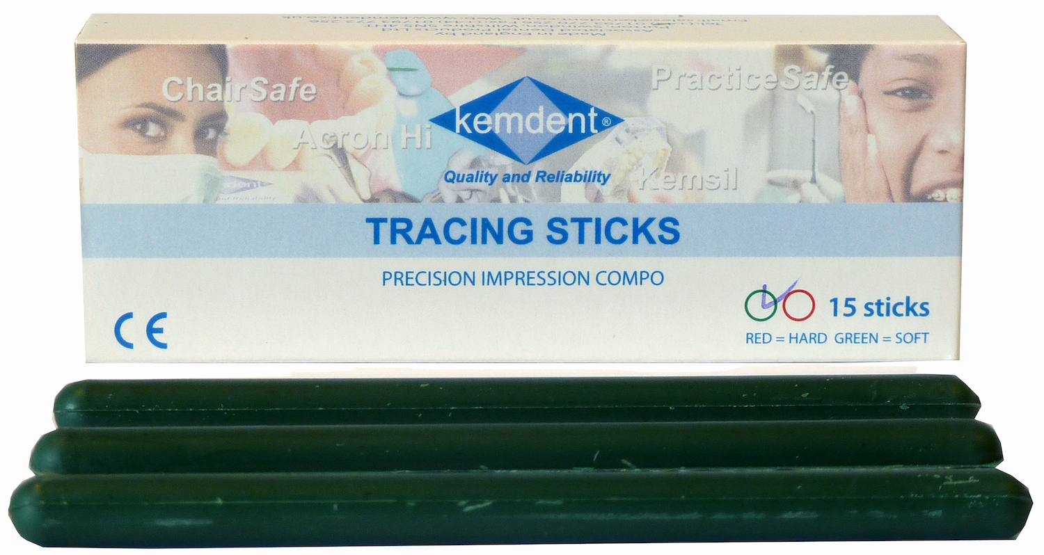
Therefore, when working with the partially edentulous patient, a conventional impression method may be advantageous to maximise the detail that can be attained. However, doing so also relies on an effective material choice. Kemdent offers clinicians a variety of exceptional impression compound materials, including the Precision Impression Compound Tracing Sticks. These are exceptional for obtaining a greater definition in the full sulcal depth or creating border extensions on impression trays. With high viscosity they can deliver exact and minute details, without compromise.
Creating comfortable, aesthetic and functional solutions for the partially edentulous patient requires an effective initial impression. Whilst digital techniques are still evolving, conventional methods may yet be preferred to avoid a wider potential variation in results when treating larger defects or working with sub-gingival margins.
Choosing when to use such solutions and effectively employ techniques such as gingival retraction, where they are suitable, is a skill in itself for clinicians. But with confidence in their materials and tools, it may be easier to attain a brilliant impression, and a fantastic final restoration, with each passing treatment.

For more information about the leading solutions available from Kemdent, please visit
www.kemdent.co.uk or call 01793 770 256

[i] Waldecker, M., Rues, S., Awounvo Awounvo, J. S., Rammelsberg, P., & Bömicke, W. (2022). In vitro accuracy of digital and conventional impressions in the partially edentulous maxilla. Clinical Oral Investigations, 26(11), 6491-6502.
[ii] Siqueira, R., Galli, M., Chen, Z., Mendonça, G., Meirelles, L., Wang, H. L., & Chan, H. L. (2021). Intraoral scanning reduces procedure time and improves patient comfort in fixed prosthodontics and implant dentistry: a systematic review. Clinical oral investigations, 1-15.
[iii] Kumar, L., Mattoo, K. A., Jain, S., Khalid, I., Kota, M. Z., Baig, F. A., … & Kanji, M. A. (2023). A Clinical Study of 50 Partially Edentulous Patients with Fixed Partial Denture Restorations to Compare Clinical Parameters and Changes in Gingival Sulcus Width After Displacement with 2 Different Gingival Retraction Cord Materials (Cotton and Polymer). Medical Science Monitor: International Medical Journal of Experimental and Clinical Research, 29, e940098-1.
[iv] Ferrari Cagidiaco, E., Zarone, F., Discepoli, N., Joda, T., & Ferrari, M. (2021). Analysis of the reproducibility of subgingival vertical margins using intraoral optical scanning (IOS): a randomized controlled pilot trial. Journal of Clinical Medicine, 10(5), 941.