Maxillary sinus exposure after tooth extraction is a common occurrence that requires prompt identification
Maxillary sinus exposure after tooth extraction refers to the accidental perforation or opening of the maxillary sinus during the process of removing a tooth from the upper jaw. This can occur due to various reasons, such as a thin bone wall separating the sinus from the tooth socket or excessive force applied during the extraction.[i]
As you know, the maxillary sinus is a cavity located in the cheeks, above the upper teeth.[ii] It is lined with mucous membranes and serves to filter, humidify, and warm the air we breathe. When a tooth is extracted from the upper jaw, there is the risk of creating a communication inadvertently between the oral cavity and the maxillary sinus.
To manage maxillary sinus exposure, prompt identification and appropriate treatment are crucial.[iii] If suspected during the extraction, the dentist may stop the procedure and assess the extent of the exposure. A visual inspection and radiographic imaging, such as a periapical or panoramic x-ray, can help determine the size, location, and severity of the sinus opening.[iv]
The classification of maxillary sinus exposure can vary, but it generally includes three categories: small, moderate, and large. A small exposure refers to a perforation measuring less than 2-3mm in diameter, a moderate exposure ranges between 3-5mm, and a large exposure exceeds 5mm.i
Simple procedure
Immediate management of small exposures usually involves the simple procedure of primary closure, suturing the perforation site with resorbable or non-resorbable sutures. It is essential to inform the patient about potential complications, such as sinus infection, during the follow-up period.[v]
In cases of moderate or large exposures, primary closure may not be feasible due to the size of the perforation or compromised soft tissue. In these situations, a secondary closure technique would be employed. The secondary closure involves creating a surgical or buccal advancement flap to access and close the sinus communication. This technique allows direct visualisation and proper manipulation of the exposed sinus membrane.[vi]
In some instances, the dentist may decide not to close the maxillary sinus exposure immediately. This may be due to factors such as inflammation, infection, or inadequate visualisation of the perforation site. In such cases, the clinician may choose to place a resorbable or non-resorbable membrane over the perforation site to act as a barrier and promote healing. This guided tissue regeneration technique allows the soft tissue to regenerate, closing the communication between the sinus and the oral cavity over time.iii
Complications
Post-operative care is crucial for patients who have experienced maxillary sinus exposure during tooth extraction. Dentists may prescribe antibiotics, nasal decongestants, or saline rinses to minimise the risk of infection and promote healing. Following the dental professional’s instructions regarding diet, oral hygiene, and activity restrictions is essential to prevent complications.[vii]
Complications that can arise from maxillary sinus exposure include sinusitis, oroantral fistula, sinus infection, implant failure, and delayed healing.iii Patients should be informed about these potential risks and encouraged to report any persistent symptoms, such as pain, swelling, discharge, or difficulty in breathing through the nose.
Given that a tooth extraction is one of the treatments most feared by many patients, using the correct – and best – instruments is vital for both the treatment and the patient experience.[viii]
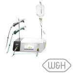
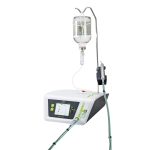
With Piezomed from W&H, dentists and oral surgeons have all the advantages of cutting-edge ultrasound technology at their fingertips. Ideal for extractions, its high frequency microvibrations allow for incredibly precise surgical procedures – only bone substance is resected – and its cavitation effect ensures an almost blood-free surgical site. The surrounding soft tissue remains largely intact, which means less pain for the patient and a quicker healing time. As soon as an instrument is inserted, Piezomed automatically detects the instrument being used and assigns it to the correct power class. This not only facilitates operation but also increases safety in oral surgery, including maxillary sinus exposure corrections.
Maxillary sinus exposure after tooth extraction is a relatively common occurrence that requires prompt identification and appropriate management. The classification of the exposure determines the treatment approach, which may range from primary closure to guided tissue regeneration or surgical flap techniques. Close monitoring and post-operative care are essential to minimise the risk of complications and ensure optimal healing.

To find out more visit www.wh.com/en_uk, call 01727 874990 or email
office.uk@wh.com

[i] Somayaji, K., Muliya, V.S., KG, M.R. et al. A literature review of the maxillary sinus with special emphasis on its anatomy and odontogenic diseases associated with it. Egypt J Otolaryngol 39, 173 (2023). https://doi.org/10.1186/s43163-023-00536-7 [Accessed January 2024]
[ii] Whyte A, Boeddinghaus R. Correction to The maxillary sinus: physiology, development and imaging anatomy. Dentomaxillofac Radiol. 2019 Dec;48(8):20190205c. doi: 10.1259/dmfr.20190205.c. Epub 2019 Sep 10. Erratum for: Dentomaxillofac Radiol. 2019 Dec;48(8):20190205. PMID: 31502867; PMCID: PMC6951093. [Accessed January 2024]
[iii] Khandelwal P, Hajira N. Management of Oro-antral Communication and Fistula: Various Surgical Options. World J Plast Surg. 2017 Jan;6(1):3-8. PMID: 28289607; PMCID: PMC5339603. [Accessed January 2024]
[iv] Vestin Fredriksson, M., Kuoljok, J., Flygare, L., Berggren, D., Tano, K. (2019) Clinical Manifestations and Symptoms of Maxillary Sinusitis of Odontogenic Origin Demonstrated by Cone Beam Computed Tomography Journal of General Practice, 7(1): 371 https://doi.org/10.4172/2329-9126.1000371 [Accessed January 2024]
[v] Parvini P, Obreja K, Begic A, Schwarz F, Becker J, Sader R, Salti L. Decision-making in closure of oroantral communication and fistula. Int J Implant Dent. 2019 Apr 1;5(1):13. doi: 10.1186/s40729-019-0165-7. PMID: 30931487; PMCID: PMC6441669. [Accessed January 2024]
[vi] Belmehdi A, El Harti K. Management of oroantral communication using buccal advanced flap. Pan Afr Med J. 2019 Oct 3;34:69. doi: 10.11604/pamj.2019.34.69.19959. PMID: 31819785; PMCID: PMC6884724. [Accessed January 2024]
[vii] Testori T, Drago L, Wallace SS, Capelli M, Galli F, Zuffetti F, Parenti A, Deflorian M, Fumagalli L, Weinstein RL, Maiorana C, Di Stefano D, Valentini P, Giannì AB, Chiapasco M, Vinci R, Pignataro L, Mantovani M, Torretta S, Pipolo C, Felisati G, Padoan G, Castelnuovo P, Mattina R, Del Fabbro M. Prevention and treatment of postoperative infections after sinus elevation surgery: clinical consensus and recommendations. Int J Dent. 2012;2012:365809. doi: 10.1155/2012/365809. Epub 2012 Aug 9. PMID: 22927851; PMCID: PMC3423929. [Accessed January 2024]
[viii] Dhanraj Ganapathy, Sivesh Sangar, Hemavathy Muralidoss. Fear Of Dental Extraction. Int J Dentistr y Oral Sci. 2021;08(05):2470-2473 https://www.researchgate.net/publication/352253205_Fear_Of_Dental_Extraction