

3D imaging in daily dentistry
Featured Products Promotional FeaturesPosted by: Dental Design 26th July 2023

Imaging has become crucial in many aspects of dentistry. Used to assist dentists in diagnosis and treatment planning, radiographs allow clinicians to gain a clearer view of the patient’s anatomy, and pick up on issues which may have otherwise gone unnoticed. 3D imaging, in particular, provides clinicians with precise information about patients’ conditions, making it an especially valuable tool in day-to-day dentistry. With more practices across the country implementing digital dental solutions, it is useful to understand where dental imaging technology began, and the many ways it can be used routinely in practice.
The introduction of 3D imaging to dentistry
When Wilhelm Röntgen discovered X-rays in 1895, he kickstarted the development of a valuable tool in the medical and dental professions. In the months and years after his discovery, technology was further explored by pioneers at the time, developing to make exposures of bone fractures and bullets in injured soldiers, in addition to early attempts at radiotherapy. Just two weeks after Röntgen’s discovery, the first dental radiograph was taken, and in 1896, the first dental X-ray laboratory was established.[i] Many further innovations have been made since then, leading to the development of the cone beam computed tomography (CBCT) scanner in the 1990s, which is now the conventional 3D dental imaging method.i
Benefits in day-to-day dentistry
 CBCT is an incredibly useful imaging method in daily dentistry. This 3D imaging method allows clinicians to visualise and assess the patient’s bony anatomical structures and any pathologies with accuracy thanks to its high diagnostic precision.[ii] CBCT imaging is particularly useful when 2D imaging methods don’t provide enough information needed for accurate diagnosis and treatment planning, and should be used where clinically necessary, to acquire a clear picture of the patient’s anatomy.ii
When compared to other imaging methods, such as CT and MRI imaging, CBCT is preferred for diagnosing a number of conditions because of its low radiation capabilities, high-resolution images, wide ranging fields of view, and value for money.[iii] The use of 3D imaging should be considered when needed to achieve an accurate diagnosis, and when required for treatment planning. Another way that CBCT imaging is used as part of day-to-day workflows, is for follow-up examinations. For example, in many cases, clinical examinations and 2D radiographs are not sufficient to establish healing. Should this be the case, CBCT imaging could be appropriate to determine the patient’s condition with accuracy.iii
Enhance communication
In addition to its benefits as a tool for diagnosis and treatment planning, CBCT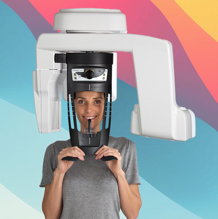 imaging acts as a useful communication tool both amongst the dental team, and with patients. Because images obtained using CBCT produce comprehensive descriptions of the patient’s anatomy, they facilitate communication between clinicians, specialists, and dental technicians. This means that should a patient require a dental implant, for example, the dental team are able to work together to fabricate an ideal prosthesis based on surgical guides derived from CBCT images.[iv] By using CBCT images as a tool for treatment planning, the dental team can expect to produce more accurate results in all fields.
3D imaging is also particularly useful when communicating with patients. The comprehensive view of their dental anatomy makes it simpler for them to understand their diagnosis, as well as the reality of any treatment plans which you might suggest. With the implementation of digital treatment planning in many dental practices, the possibilities for communication expand further, as patients will be able to see a virtual model of their anatomy which includes their hard and soft tissues.[v] By providing patients with these additional communication tools, you are likely to see a boost in treatment acceptance and patient satisfaction, with patients feeling confident in your abilities, and experiencing a more in depth education about their own oral health.
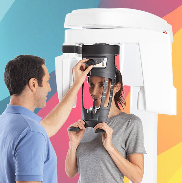
The CS 8100 3D Evo Edition CBCT scanner from Carestream Dental is the ideal solution for any dental practice. With the ability to produce high-resolution 3D images with limited artifacts, the CS 8100 is trusted by thousands of general dentists and endodontists around the world. Plus, its award-winning design is able to fit into tight spaces, perfect even for smaller dental practices.
CBCT imaging is becoming more widely implemented into many practices’ digital workflows, with clinicians and patients enjoying the numerous benefits it brings. Imaging solutions are constantly being developed, with options suitable for smaller dental practices, making this incredibly useful technology more easily accessible for use in daily dentistry.
![]()
For more information on Carestream Dental visit www.carestreamdental.co.uk
For the latest news and updates, follow us on Facebook and Instagram @carestreamdental.uk
Nimisha Nariapara
Bio
Nimisha is the Trade Marketing Manager at Carestream Dental covering the UK, Middle East, Nordics, South Africa, Russia and CIS regions. She has worked at Carestream Dental for the past 7 years, where she has developed her marketing skills and industry knowledge to bring the core values and philosophy of the company to the market.
Kind regards
[i] Pauwels, R. “History of dental radiography: Evolution of 2D and 3D imaging modalities.” Med Phys Int 8.1 (2020): 235-77.
[ii] Hung, Kuofeng, et al. “Current applications, opportunities, and limitations of AI for 3D imaging in dental research and practice.” International Journal of Environmental Research and Public Health 17.12 (2020): 4424.
[iii] Cohenca, Nestor, and Adrian Silberman. “Contemporary imaging for the diagnosis and treatment of traumatic dental injuries: A review.” Dental Traumatology 33.5 (2017): 321-328.
[iv] Rios, Hector F., Wenche S. Borgnakke, and Erika Benavides. “The use of cone‐beam computed tomography in management of patients requiring dental implants: an American Academy of Periodontology best evidence review.” Journal of periodontology 88.10 (2017): 946-959.
[v] Swennen, Gwen RJ, Wouter Mollemans, and Filip Schutyser. “Three-dimensional treatment planning of orthognathic surgery in the era of virtual imaging.” Journal of oral and maxillofacial surgery 67.10 (2009): 2080-2092.


