

A challenging endo case: the difficulties posed when lower premolars have multiple canals
Featured Products Promotional FeaturesPosted by: Dental Design 24th November 2022

Dr Sagi Shavit graduated from dental school in 2009 in Bucharest and shortly afterwards developed a special interest in endodontics. He undertook a number of endodontic training courses, including microsurgical endodontics, in London and in 2015, he gained an MSc in Endodontics from the University of Chester. He now treats endodontic patients exclusively at Cleveland Terrace Dental Practice, Darlington.
Here he presents a case where complex anatomy meant challenges for the entire treatment sequence.
Background, presentation and diagnosis
A female patient in her late 20s was referred to our clinic regarding her lower premolar.
She complained of occasional constant painful episodes and pain upon mastication. A clinical examination revealed that the tooth was not restored or carious, but seemed slightly discoloured and was not responsive to cold tests. It was also tender to vertical pressure.
The patient had orthodontic treatment several years ago and there was no recollection of trauma.
Radiographic examination (see figure 1) shows a single premolar, normal bone levels and discrete widening of the apical periodontal space. Further careful examination of the preoperative radiograph shows that there is a single wide canal which abruptly disappears mid-root. This is an indication of a separation of this canal into two separate canals.
Based upon the clinical and radiographic findings, a diagnosis of chronic apical periodontitis was placed. The treatment options of endodontic treatment or extraction of the tooth were discussed, and the patient decided to have endodontic treatment.
Treatment pathway
The tooth was accessed under rubber dam and the canals were located with the aid of the dental operating microscope (see figure 2).
The access to the two canals was refined using the HyFlex™ EDM Orifice Opener (25/.12) from COLTENE, in order to allow better access into each canal.
The working length was determined using the CanalPro™ electronic Apex Locator, also available from COLTENE, and verified with a radiograph (see figure 3). This also allowed me to understand the exact anatomy of the root canal system, which was proved to be type II Weine.
A smooth glide path was achieved using manual 8 and 10 K files, and the 10/.05 GlidePath file from the HyFlex™ EDM system.
Mechanical instrumentation continued using the 20/04 in both canals and the HyFlex™ EDM OneFile in the buccal canal, which was the main canal. This was done to avoid over instrumentation of the canals, which may lead to iatrogenic accidents.
This was performed under copious irrigation of 5.25% sodium hypochlorite, and a final rise with 17% EDTA. Both irrigation solutions were activated ultrasonically using the cordless CanalPro™ Endo Ultra Irrigation Activator.
The gutta percha points were fitted inside the wet canals to verify ‘tug-back’ and another radiograph was acquired to confirm fit (see figure 4). The canals were then dried using matching paper points, and obturated using warm vertical compaction (see figure 5 and figure 6).
The access cavity was sealed with resin composite and the patient discharged back to their referring dentist. The postoperative radiograph (see figure 7) shows adequate three-dimensional obturation of the endodontic space.
Conclusions
Lower premolars with multiple canals pose a challenge throughout the entire endodontic treatment, from diagnosis and treatment planning, through access and instrumentation, to proper irrigation and obturation.
COLTENE provides a range of high-quality endodontic tools, materials and equipment that allow us, as clinicians, to provide the best treatments to our patients. HyFlex™ EDM files are super elastic, efficient and safe. These files will own any canal you’ll give them. The CanalPro™ Apex Locator provides crisp and accurate readings of the working length and is indispensable tool during endodontic treatment.
Irrigation, the most important part of the endodontic treatment, is much more efficient using ultrasonic devices, such as the CanalPro™ Endo Ultra. The matching HyFlex™ EDM paper points and gutta percha points allow safe and accurate obturation of the endodontic space.
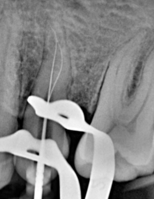
Figure 1. Pre-op radiograph
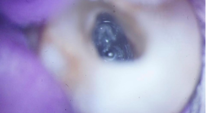
Figure 2. Access cavity and view of the two canals
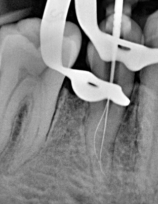
Figure 3. Working length radiograph
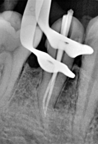
Figure 4. Cone fit radiograph
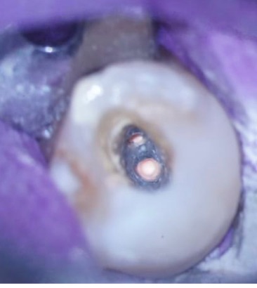
Figure 5. View of both canals obturated
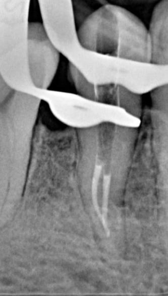
Figure 6. ‘Down-pack’ radiograph
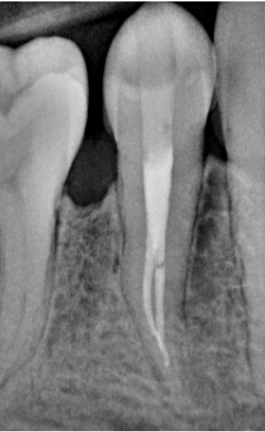
Figure 7. Post operative radiograph
For more on COLTENE, visit www.coltene.com,
email info.uk@coltene.com or call 0800 254 5115.


