Focus on human factors behind incidents

Leo Briggs, deputy head of the DDU examines how we need to better understand human error when looking at improving patient safety.

Human error is inevitable in any walk of life. But in dentistry, where one slip has the potential to cause lasting harm to a patient, the stakes are higher. In an ideal world, every conscientious dental professional would have the time, space and support to treat patients. They would never be fatigued, under time pressure because of a full waiting room, their usual dental nurse would always be in attendance, equipment would operate perfectly and every patient would be calm personified.

Of course, the reality of clinical practice is far from perfect and factors like tiredness or poor team communication can contribute to human error. Sadly, when adverse incidents do occur, dental professionals may be individually blamed or face an investigation – when it would be better for everyone to find out what went wrong, and to learn from it.

The role of human factors is to examine why errors occur and then develop strategies to ‘absorb’ the risk by improving work systems, processes and technological innovation. In an interview in the DDU journal, Professor Simon Wright, chairman of the National Advisory Board for Human Factors in Dentistry explained: “The first thing we have to do is create a culture within dentistry where we all understand that we make errors and learn from them, rather than try to hide the fact we are making them because of the possible consequences.”

But how can we do this in practice? It takes a team effort as the following fictitious case shows.

A foundation dentist had been asked by the orthodontist treating a 14-year old girl to remove a retained lower right deciduous second molar (LRE). When the foundation dentist examined the patient, he incorrectly identified the lower right first permanent molar (LR6) as the tooth to be removed.

After administering local anaesthetic and checking for adequate analgesia, the dentist prepared to extract the tooth. Fortunately, the dental nurse was observing closely and as the dentist was about to begin the extraction the nurse intervened to point out the error.

The dentist paused the procedure, and identified the correct tooth by checking it in the mouth against the referral letter from the orthodontist, the dental charting and radiographs, and double-checked it with the patient, the accompanying parent, and the dental nurse. Having satisfied himself that the correct tooth had now been positively identified, the LRE was removed uneventfully.

The foundation dentist discussed this ‘near miss’ with his educational supervisor, and the nurse was praised for intervening and speaking up to avoid what would otherwise have been a serious adverse incident.

A root cause analysis was carried out and there was a discussion about the near miss at a practice meeting. One of the outcomes was that in the future the practice would adopt the Royal College of Surgeons dental LocSSIP tool aimed at preventing erroneous extractions.

You can see more about this issue in the latest issue of the DDU journal ddujournal.theddu.com

National Dental Nursing Conference

The British Dental Conference and Dentistry Show 2020 will offer an array of learning and networking opportunities for all dental nurses.

Ensuring the relevance and quality of the educational programme, the British Association of Dental Nurses (BADN) will be hosting the National Dental Nursing Conference as part of the event. The Dental Nurses Forum will be chaired by the BADN and will see leading speakers share their wealth of expertise and advice with delegates, covering everything from patient communication and management to skill development and career progression.

Plus, BADN members will be able to relax in the BADN Lounge between lectures and exhibition browsing on stand L95.

For all this and much more, register for your free delegate pass online today!


The British Dental Conference and Dentistry Show 2020 – 15th and 16th May –Birmingham NEC, co-located with DTS.

For all the latest information, visit www.thedentistryshow.co.uk, call 020 7348 5270 or email dentistry@closerstillmedia.com

Helpful advice whenever needed

“As I was buying a practice through Dental Elite, it was convenient to also use their dedicated CQC service.” 

Dr Mani Shakibpour shares his experience of his CQC application during a practice acquisition with Dental Elite. He continues:

“The interview was the most challenging aspect of the CQC application as I didn’t know what to expect. Dental Elite provided guidance on what might be asked and helped me to prepare.

“I was kept up-to-date on progress throughout and the team were very easy to reach if I had any queries. I was very pleased with the help both Helen [Craine] and Bailey [Rowles] provided – they gave great advice whenever it was necessary. As such, I would recommend the CQC service from Dental Elite to other dentists.”


For more information contact Dental Elite. Visit www.dentalelite.co.uk, email info@dentalelite.co.uk or call 01788 545 900

More than just teeth

November was Mouth Cancer Action month – your practice may have posted ‘blue lip selfies’ on social media to show its support. Since its creation, the annual awareness campaign has grown considerably and this has been driven by necessity. Over 8,000 new cases of mouth and oropharyngeal cancer are diagnosed in the UK every year and numbers continue to rise.[i] Mouth Cancer Action Month is well supported across the profession. There is a comprehensive range of resources to help patients recognise the signs and symptoms to look for and the importance of early diagnosis.

Of course, mouth cancer awareness is a year-round issue. It brings together key elements at the heart of prevention; good lifestyle choices, regular dental appointments, thorough oral hygiene and patients being alert to any changes in their oral cavity. This is perhaps what is so frustrating about the figures for mouth and oropharyngeal cancers; the message is clear and simple, yet new cases are increasing.

Practising better preventive care, every day, must be at the top of patients’ New Year’s Resolutions lists to reduce the risk of dental problems, including cancer of the mouth, head and neck, as well as a host of other conditions. Dental care professionals need
to find new ways to encourage people to make long-term changes and learn to think differently about what ‘oral health’ actually means – because it’s way more than just clean teeth.

If you asked your patients what purpose a dental check-up serves, most answers would be tooth-related, such as “to see if I need a filling”, or “to find out what’s causing my toothache”. But true prevention is about getting people to understand how to keep the whole mouth healthy – the gums, cheeks, tongue and lips as well as head and neck. Regular appointments give people a time to talk about anything and everything that relates to the mouth, head and neck. Sore lips? Recurring ulcers? Pain when swallowing? These are things that should be discussed to find a solution or to decide if further investigation is required.

Rather like breast checks, patients should be shown how to check their mouth at home, so they can quickly see and feel if something is amiss. As part of Mouth Cancer Action month, there are practice resources that show people how to conduct a thorough self-examination; self-assessment should be recapped at every preventive-maintenance appointment and include a demonstration too.

Good prevention is helping patients understand how all the components of a preventive approach rely on each other. The impact of poor lifestyle choices, like drinking too much alcohol, won’t be diminished by good toothbrushing and using an expensive mouth rinse. Regularly drinking more than the recommended alcohol limit is a risk factor for seven types of cancer, including mouth, pharyngeal, oesophageal and laryngeal cancer.[ii] When combined with smoking, the risk is increased further.[iii] Eating a balanced diet including plenty of fruit and vegetables will also reduce the risk of mouth cancer plus a host of other systemic and serious diseases. These are known health messages of course, but the advantage that a DCP has over a GP is they will be talking with patients when they are feeling well. Preventive-maintenance appointments should start with a discussion of the ‘basics’ like alcohol limits, diet and smoking cessation – with referrals made if required.

Another topic that can be discussed is the importance of the HPV vaccine, for teen patients and parents bringing their teenagers to the practice. The ‘anti-vaxx’ movement has grown in profile; there is great deal of misinformation on social media about the safety of vaccines which DCPs can talk through with worried patients. Some cancers of the head and neck are caused by the HPV virus and the vaccination is now rolled out for all Year 8s.

For patients diagnosed with a form of mouth cancer, effective cleaning is an essential part of their daily routine. However, due to their cancer treatment they might find this uncomfortable or difficult. You can consider adding new tools to their home care, especially softer, gentler aids. The TANDEX UltraSoft range has solutions including toothbrushes and FLEXI™ UltraSoft interdental brushes, which are gentle on gums and designed specifically for use after surgery.

Mouth and oropharyngeal cancer rates are rising and motivating people in good behaviours to reduce their risk should be a priority all-year round. For dental care professionals, it’s about encouraging a shift in patients’ thinking. Oral health is about far more than teeth and toothbrushing. It’s about being aware of the whole mouth and how the different components of preventive care interact. Patients may be more receptive if instructions and advice are given in a less ‘formal’ setting, by a dental hygienist or dental therapist. With so much to do to improve the nation’s oral health and cancer rates, practices should grab any opportunity for the whole team to get involved in supporting patients.


For more information on Tandex’s range of products,
visit
www.tandex.dk or visit Facebook


[i] Oral Health Foundation. The State of Mouth Cancer UK Report 2018/2019. Link: https://www.dentalhealth.org/stateofmouthcancer (accessed October 2019).

[ii] Cancer Research UK. Does alcohol cause cancer? Link: https://www.cancerresearchuk.org/about-cancer/causes-of-cancer/alcohol-and-cancer/does-alcohol-cause-cancer#alcohol10  (accessed October 2019).

[iii] Cancer Research UK. Does alcohol cause cancer?

Selecting implant techniques

For many patients dental implants are – aesthetically and functionally – the best means available to treat edentulism. Success rates of 90-95% after a decade are commonly reported. However, as with any surgical procedure, complications do remain a possibility. Host factors, the implant site, surgical method and the design of the implant, can all have a bearing on the success of treatment.[i]

Many complications can be avoided through careful case selection and preoperative planning. The general health and behaviours of the patient can be a risk factor, as is the case for smokers, diabetic patients and those with epilepsy, among others. There are also aspects of a patient’s medical history that may present an absolute contraindication to treatment, such as strong immunosuppression after an organ transplant.i These and other host factors can be easily screened for.

Localised anatomical factors can influence the success of procedures and increase the potential for complications. Numerous surgical techniques and implant systems have been developed in response to particular challenges that can arise from specific anatomical factors.

Bone tissue

In many cases the primary limiting factor concerning dental implants is the quality and quantity of bone in the region. Once a permanent tooth is extracted or lost, bone resorption is a biological inevitability. Bone structure is adaptive and remodels in response to strain; the absence of a tooth reduces strain on the area beneath it, leading to reductions in the buccolingual and apicocoronal dimensions of the alveolar ridge.[ii] In addition to the normal bone remodelling process, periodontal disease and other conditions can result in inadequate bone tissue.

The insertion of a titanium implant results in increased stiffness in the mandible, making it more resistant to compressive force. Theoretically, this results in a reduction in the forces the underlying bone is subject to, and can therefore result in bone resorption continuing (if not to the extent that an empty socket causes). This can be compensated for by using implants with suitably designed retention elements, which confer the needed routine strain to maintain bone mass.ii

Where the bone is inadequate to properly support an implant, this needs to be compensated for. Methods for accomplishing this include bone augmentation using grafts and sinus lift procedures, using 3D customised implants or utilising zygomatic implants.

Among bone grafts, autografts are ideal where possible as they will not result in immunogenic complications. However, this requires additional surgery at the donor site (with the potential for morbidity there), and the amount of bone available may be quite limited. Where the patient is unsuitable for autogenous grafting, graft materials from other donors, animals, or synthetic sources are also available.[iii]

Zygomatic implants are longer than conventional dental implants, allowing the prosthesis to be anchored into the cheek bone, bypassing the need for a bone graft. However, this method can still result in complications, chiefly osseointegration failure and sinusitis.[iv]

Parts of the soft tissue will also resorb relatively quickly following extraction, including the interdental papillae. Insufficient soft tissue affects aesthetic outcomes, including the potential visibility of the implant screw. Inadequate bone will also affect how well an aesthetically pleasing gingival contour can achieved. Soft tissue augmentation can be used to restore the gingival line.[v]

Nerve tissue

The inferior alveolar nerve is a branch of the mandibular nerve that relays sensation from mandibular posterior teeth, the surrounding bone structure and the mucosa of the posterior tongue.[vi]

Many patients receiving implants already exhibit significant bone atrophy, preventing the use of long fixtures. In these instances, lateralisation of the inferior alveolar nerve (LIAN), can help provide the space required to support the prosthesis in a more ideal location and avoid injury of the nerve. LIAN requires the surgeon to expose the nerve, temporarily move it aside while the implants are placed, then permit it to fall back into position. Nerve transposition is also possible, which involves a corticotomy around the mental foramen in order to reposition it. If advanced alveolar resorption has already occurred, this procedure is contraindicated.[vii]

Biological variations in this region are not unknown, with some patients having bifid or trifid alveolar nerves. A second, or third mandibular foramen can be present, which can be observed preoperatively as a double or triple mandibular canal on a conventional panoramic radiograph. If not accounted for, these variations can lead to inadequate anaesthesia or damage to the nerve, leading to bleeding, paraesthesia or neuroma development. Where bifid or trifid mandibular canals are detected, further CBCT scanning to provide confirmation and a better view of the anatomy is advisable.[viii], [ix]

If you are dealing with a complex case or have a patient with failing implants that require complex surgical intervention, consider referring your patient to the Centre for Oral-Maxillofacial and Dental Implant Reconstruction. Led by Professor Cemal Ucer – Specialist Oral Surgeon – the practice offers a wide variety of advanced procedures, including nerve lateralisation and repositioning, allografts, and zygomatic dental implants. With a wealth of experience and state-of-the-art facilities your patient will be in the best of hands.

Inadequate bone continues to pose a challenge to providing dental implants. There are numerous techniques and technologies that can help many patients, however, successfully employing the optimal solution requires careful preoperative case-selection, knowledge and surgical skill.


Please contact Professor Ucer at ice@ucer.uk or Mel Hay at mel@mdic.co

01612 371842


[i] Raikar S., Talukdar P., Kumari S., Panda S., Oommen V., Prasad A. Factors affecting the survival rate of dental implants: a retrospective study. Journal of International Society of Preventative & Community Dentistry. 2017; 7(6): 351-355. https://www.ncbi.nlm.nih.gov/pmc/articles/PMC5774056/ October 25, 2019.

[ii] Hansson S., Halldin A. Alveolar ridge resorption after tooth extraction: a consequence of a fundamental principle of bone physiology. Journal of Dental Biomechanics. 2012; 3: 1758736012456543. https://www.ncbi.nlm.nih.gov/pmc/articles/PMC3425398/ October 25, 2019.

[iii] Raghavan R., Shajahan P., Raj J., Raju R., Monisha V., Jishnu S. Review on recent advancements of bone regeneration in dental implantology. International Journal of Applied Dental Sciences.  2018; 4(2): 161-163. http://www.oraljournal.com/archives/2018/4/2/C/4-2-29 October 25, 2019.

[iv] Molinero-Mourelle P., Baca-Gonzalez L., Gao B., Saez-Alcaide L., Helm A., Lopez-Quiles J. Surgical complications in zygomatic implants: a systematic review. Medicina Oral, Patología Oral y Cirugía Bucal. 2016; 21(6): 751-757. https://www.ncbi.nlm.nih.gov/pmc/articles/PMC5116118/ October 25, 2019.

[v] Brouwers J., Buis S., Haumann R., de Groot P., Laat B., Remijn J. Successful soft and hard tissue augmentation with platelet-rich fibrin in combination with bovine bone space maintainer in a delayed implant placement protocol in the esthetic zone: a case report.  Clinical Case Reports. 2019; 7(6): 1185-1190. https://doi.org/10.1002/ccr3.2177 October 25, 2019.

[vi] Yoon T., Robinson D., Estrin N., Tagg D., Michaud R., Dinh T. Utilization of cone beam computed tomography to determine the prevalence and anatomical characteristic of bifurcated inferior alveolar nerves. General Dentistry. 2018; 66(4): 22-26. https://www.ncbi.nlm.nih.gov/pubmed/29964244 October 25, 2019.

[vii] Abayev B., Juodzbalys G. Inferior alveolar nerve lateralization and transposition for dental implant placement. Part I: a systematic review of surgical techniques.  Journal of Oral & Maxillofacial Research. 2015; 6(1): e2. https://www.ncbi.nlm.nih.gov/pmc/articles/PMC4414233/ October 25, 2019.

[viii] Mizbah K., Gerlach N., Maal T., Bergé S., Meijer G. The clinical relevance of bifid and trifid mandibular canals. Oral and Maxillofacial Surgery. 2012; 16(1): 147-151. https://link.springer.com/article/10.1007/s10006-011-0278-5 October 25, 2019.

[ix] Kumar V. Bifid mandibular canal: an aberrant anatomic variation. Journal of Dentomaxillofacial Science. 2017; 2(2): 133-134. https://jdmfs.org/index.php/jdmfs/article/view/531 October 25, 2019.

Leading dental adhesive from the makers of Panavia V5

From the makers of Panavia V5, J&S Davis brings you Clearfil Universal Bond Quick – the Swiss army knife of adhesives!

Highly economic with up to 250 applications per 5ml bottle, Clearfil Universal Bond Quick contains Kuraray’s original MDP monomer for optimal bond strength and durability. It is also quick and easy to apply – in just 15 seconds – and is suitable for use in all etch procedures.

Further still, the low film thickness (5-10μm) ensures precision fit every time.

Contact J&S Davis direct or order through your preferred dealer today.


For more information on the industry-leading products available from J&S Davis, visit www.js-davis.co.uk, call 01438 747 344 or email jsdsales@js-davis.co.uk

Achieving optimal results in full-arch implant treatment

Dr Martin Wanendeya demonstrates through a case study how he achieved excellent aesthetics and function with an FP1 dental implant restoration in his patient’s upper arch. 


A male patient presented with several mobile teeth and a loose partial denture in his upper arch that he had retained for a while. He also had existing dental implants in the upper right region that had been placed recently.

The main problem was that the partial denture was being held in place with a cement-retained implant crown in the UR5. This meant that the implant crown kept coming off and the denture was becoming loose – the patient had already been through a cycle where the denture had detached and was reattached several times. In addition, peri-implantitis had developed on and around an implant-supported denture that had been placed in the patient’s lower arch. He was unable to chew or eat properly, so he came to the practice to explore his options.

Treatment options were discussed with the patient, which included a new denture, an anterior bridge and posterior implant, an implant-retained overdenture, or a fixed implant bridge. The patient was keen on having a fixed implant bridge, so planning was carried out to determine where the ideal tooth positions would be based on the patient’s remaining dentition and his smile.

Placement of four dental implants in the upper arch was planned for, as was treatment for peri-implantitis and removal of the implant in the lower arch. The patient also decided to see how he would get on with a new replacement denture in his lower arch. 

Treatment planning

Treatment planning involved taking an impression and using this to make a shell. This is a device that enables clinicians to visualise the tooth position and shape, as well as the gingival margin. In this case, an approach was planned where – rather than lift a flap, remove the hard and soft tissue, and provide the patient with a fixed denture (i.e. a pink and white prosthesis) – a combination of augmentation, precision placement and soft tissue manipulation would be carried out. This would ensure a result where there would be white teeth emerging from the patient’s natural gum.

Implant placement

On the day of surgery, the remaining teeth in the upper arch were extracted and the abutments removed from the existing implants. Four Ankylos® implants were placed immediately following extraction and Ankylos® Balance Base abutments fitted to all six implants in the upper arch that would support the prosthesis.

Using the WeldOne™ intraoral welding technique, a welded framework was then made and bent round the implants. This metal frame was cut, trimmed and shaped, before it was treated to become opaque. The shell was then picked up using a special handpiece that also maintained the screw-access hole in the prosthesis.

Following this, a connective tissue graft was harvested from the tuberosity and placed into the anterior region of the mouth in order to boost the soft tissue profile and improve appearance in these areas.[i] Bio-Oss® small granule was also placed into the extraction sockets so as to, again, enhance results.

A provisional bridge was then made and screwed onto the implants on the day of surgery. Sutures were placed around the UL2 and UR2 areas in order to hold the soft tissue in the ideal position, which were left to heal for two weeks.

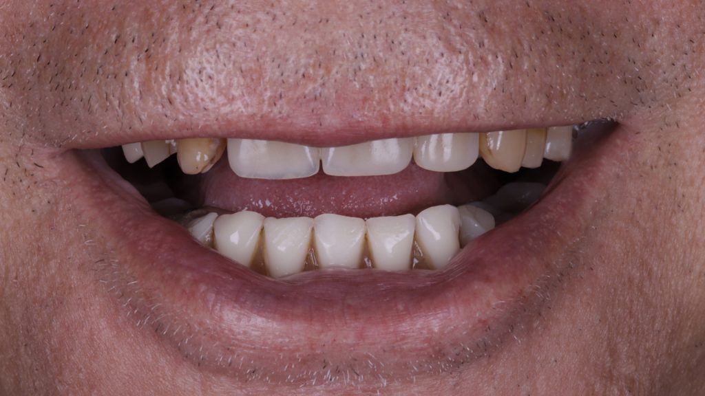
Figure 1 Full view of smile pre-implant surgery

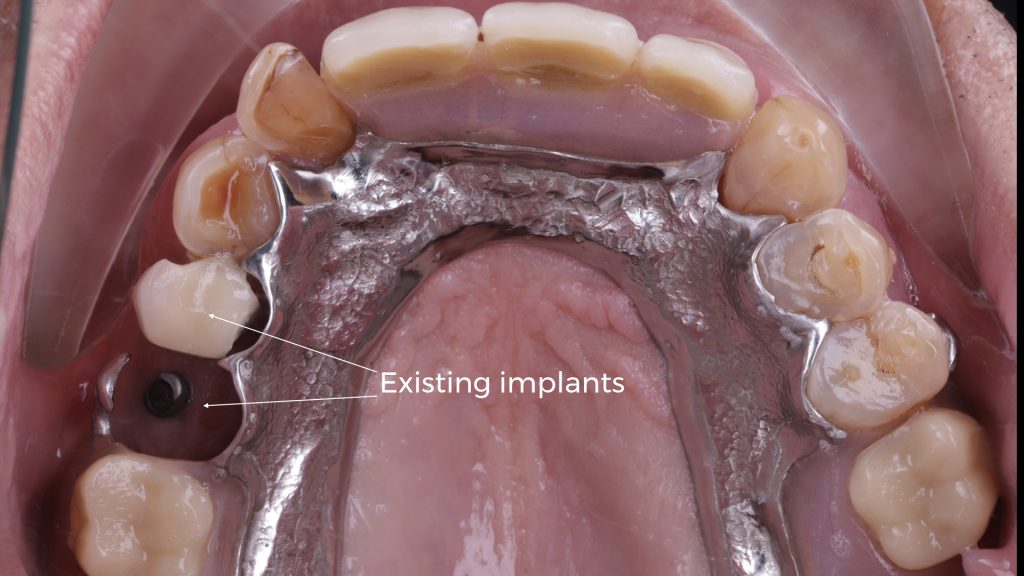
Figure 2 Upper arch with existing implants

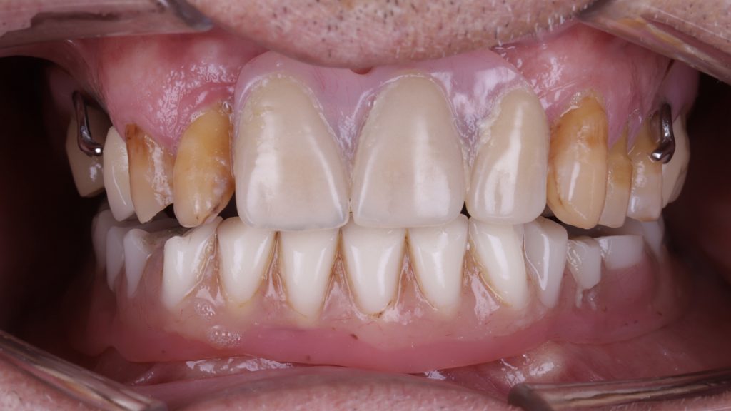
Figure 3 Dentures in lower and upper arch

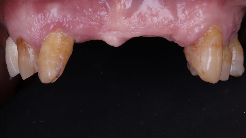
Figure 4 Denture removed in upper arch

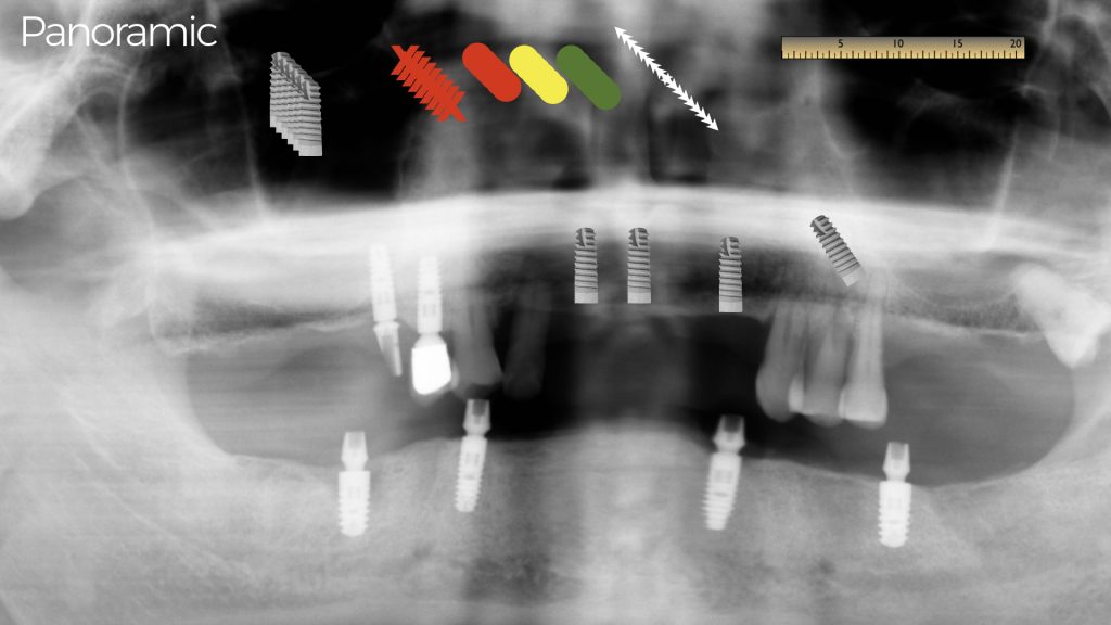
Figure 5 Implant treatment planning

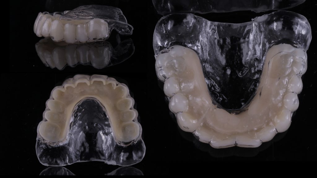
Figure 6 Shell for the temporary restoration

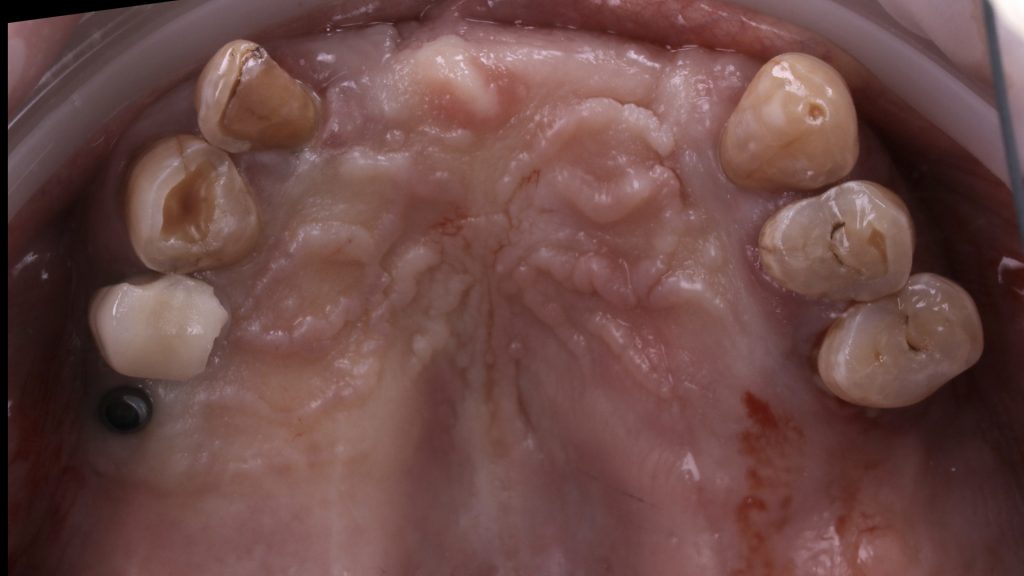
Figure 7 Upper arch pre-extraction

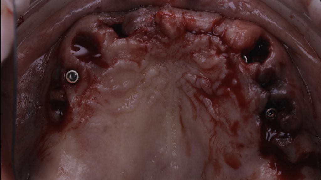
Figure 8 Implants placed in upper arch

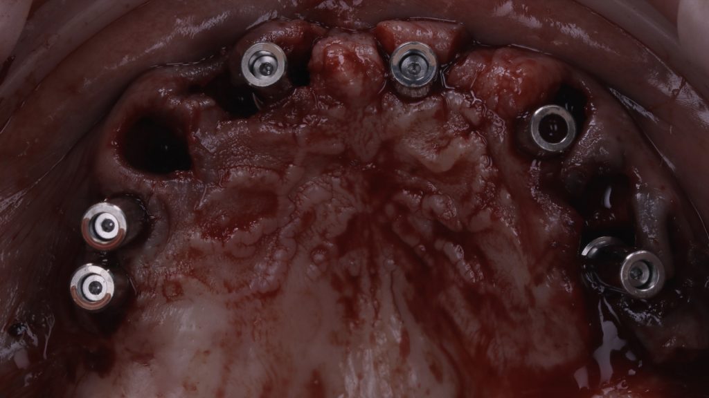
Figure 9 Implant abutments fitted

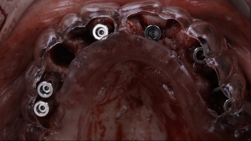
Figure 10 Try in of the prosthesis shell

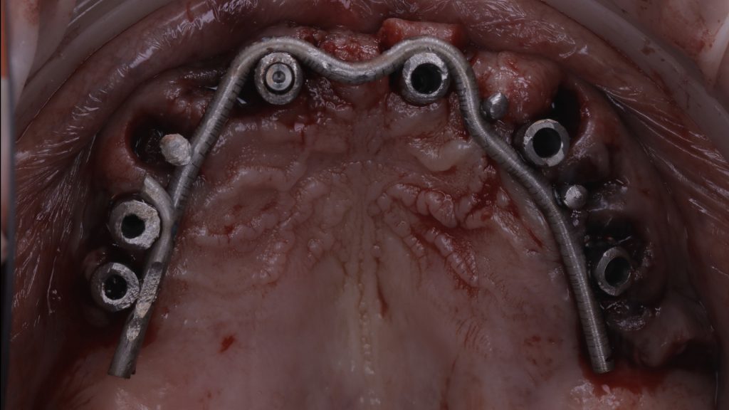
Figure 11 Intraorally welded framework

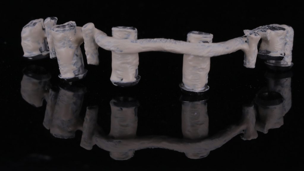
Figure 12 Opaque framework

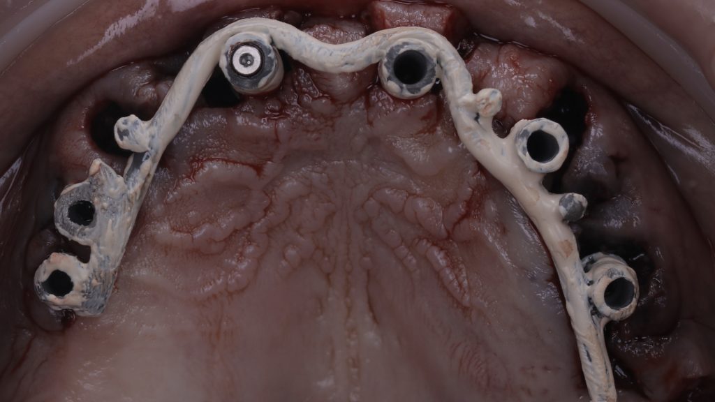
Figure 13 Try in of the opaque framework

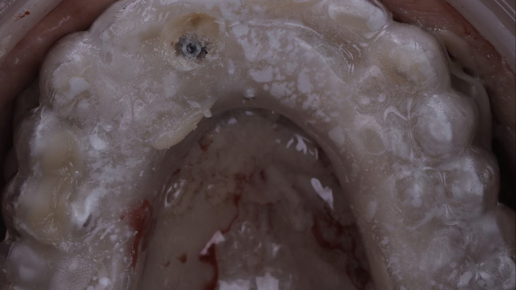
Figure 14 Try in of the prosthesis shell

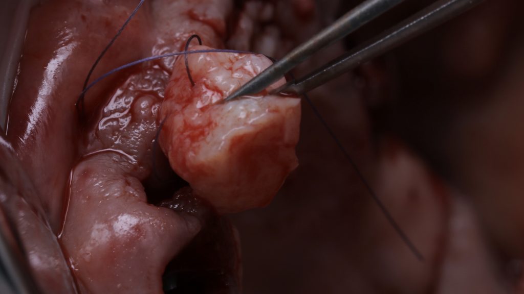
Figure 15 Suturing of soft tissue graft

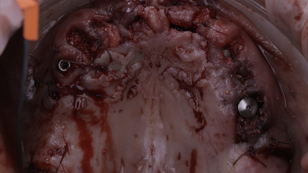
Figure 16 Soft tissue graft in the upper arch

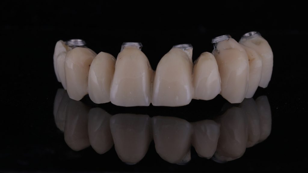
Figure 17 The provisional bridge

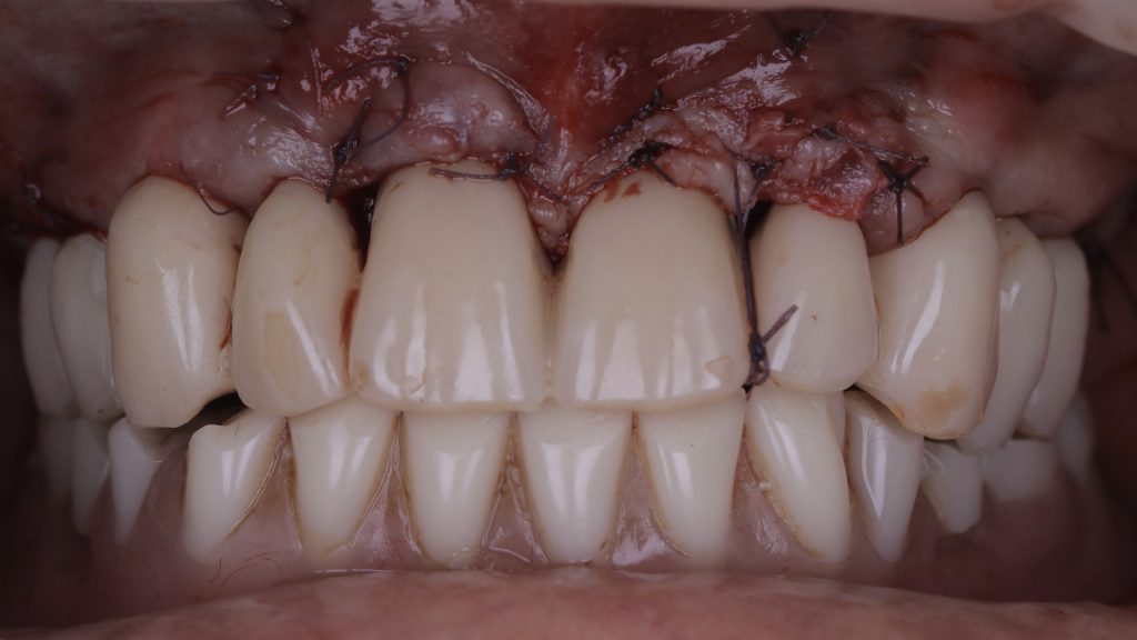
Figure 18 Temporary restoration fitted

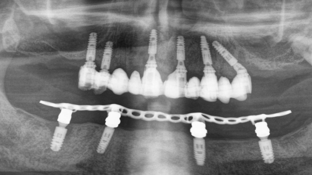
Figure 19 Radiograph following implant surgery

Implant restoration

Following approximately 3 to 4 months of further healing, the patient returned to the practice for the restorative phase of treatment. Once the patient had been assessed and found to have pink, healthy tissue, the provisional bridge was removed to prepare for the final restoration.

Elos Accurate® scan bodies were placed on each implant and a digital image taken using the TRIOS intraoral scanner, which was able to capture the soft tissue shape and implant positions. This scan was then sent to the Uniqa dental laboratory, where dental technicians – Alina and Khristo – copied the shape of the provisional restoration, produced a wax-up model and sent back a PMMA shell. Figure 28 shows the PMMA shell on the Elos® link abutments taken from the scan. This was then tried in the mouth, before aesthetics, phonetics and function were assessed.

The original intraoral scans were used to make an implant verification jig (IVJ) with a pattern resin. This was verified in the mouth and on this occasion, the IVJ fit without needing to be trimmed or shaped. Another pattern resin – primopattern from primotec – was then used to capture the soft tissue shape in the mouth. Figure 29 shows the two pattern resins in place, which were then given to the dental technicians to create a hard model.

From this model, a milled zirconia bridge was made and stained using MiYO stain. The bridge was then tried in the mouth and checked for fit, tissue shape and occlusion. The Elos® link abutments were removed and refitted. The restoration was sent back to the lab for final finishing before it was fitted. Figure 31 demonstrates excellent soft tissue integration following the final fit, while figure 32 shows both the new bridge in the upper arch and a new implant-supported denture that was fitted to the lower arch. As originally planned, the implant in the lower arch that had peri-implantitis was also removed.

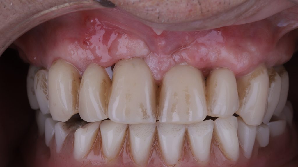
Figure 20 Soft tissue healing post-implant placement

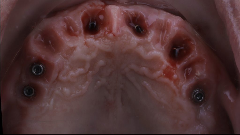
Figure 21 Provisional bridge removed

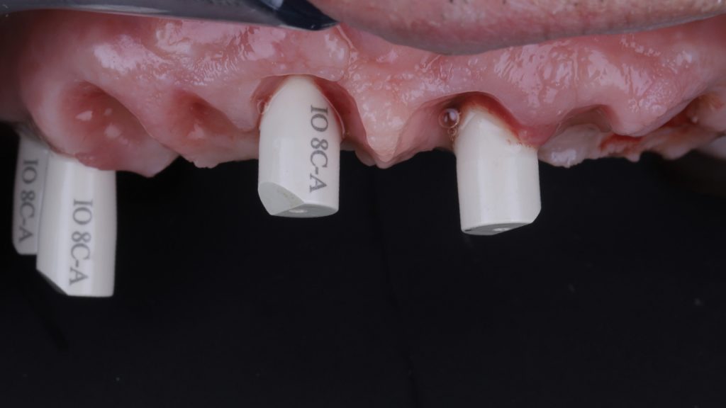
Figure 22 Scan bodies placed

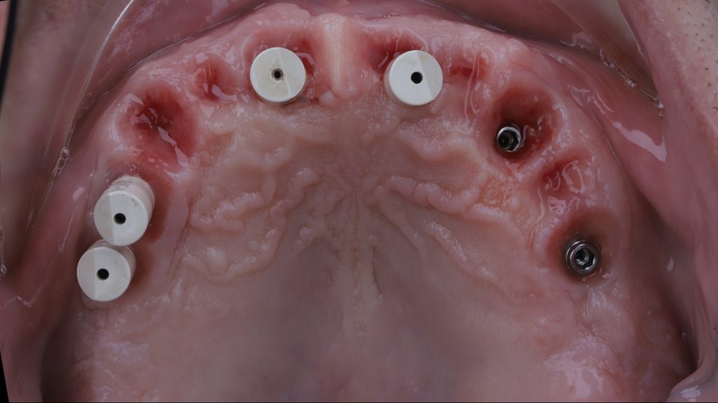
Figure 23 Upper arch with scan bodies in place

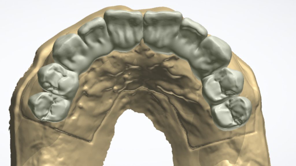
Figure 24 Intraoral scan of soft tissue and restoration

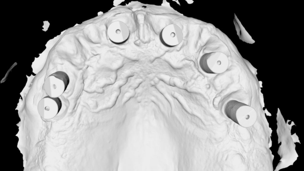
Figure 25 Intraoral scan of scan bodies in upper arch

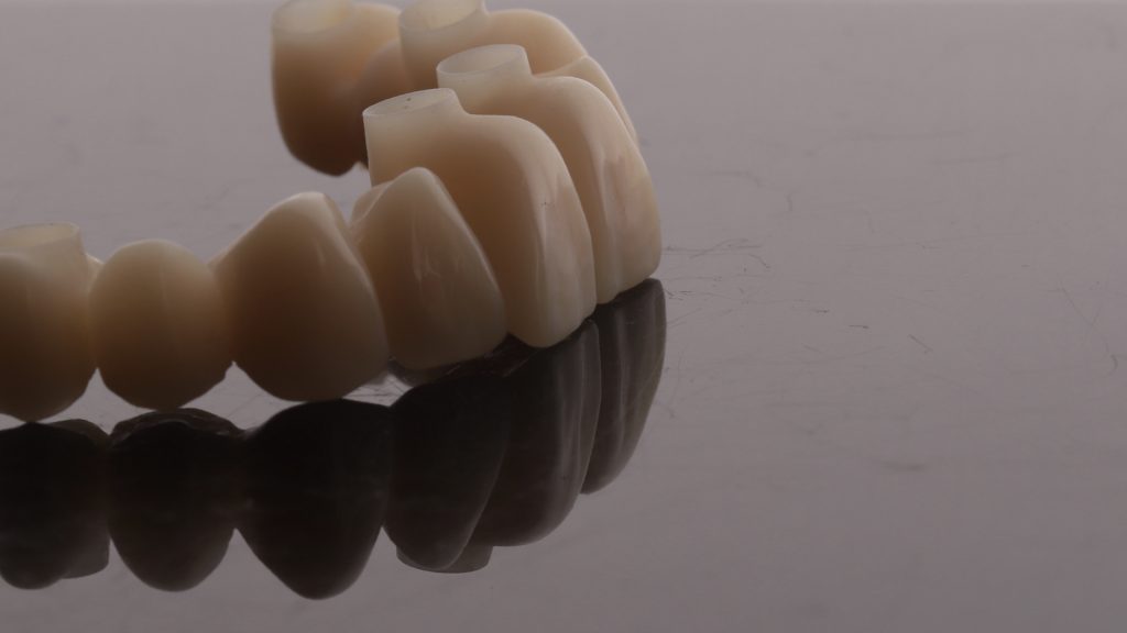
Figure 26 PMMA shell

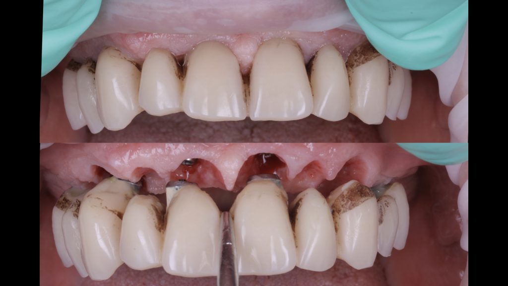
Figure 27 Removal of the temporary bridge

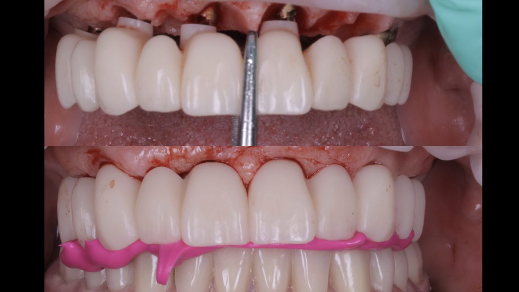
Figure 28 Try in of the PMMA shell

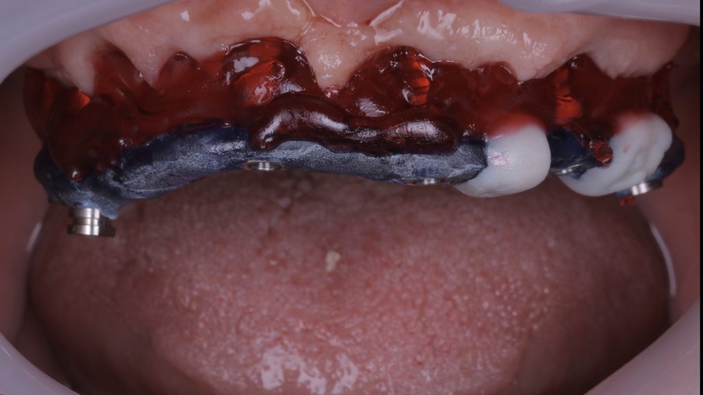
Figure 29 Pattern resins in place

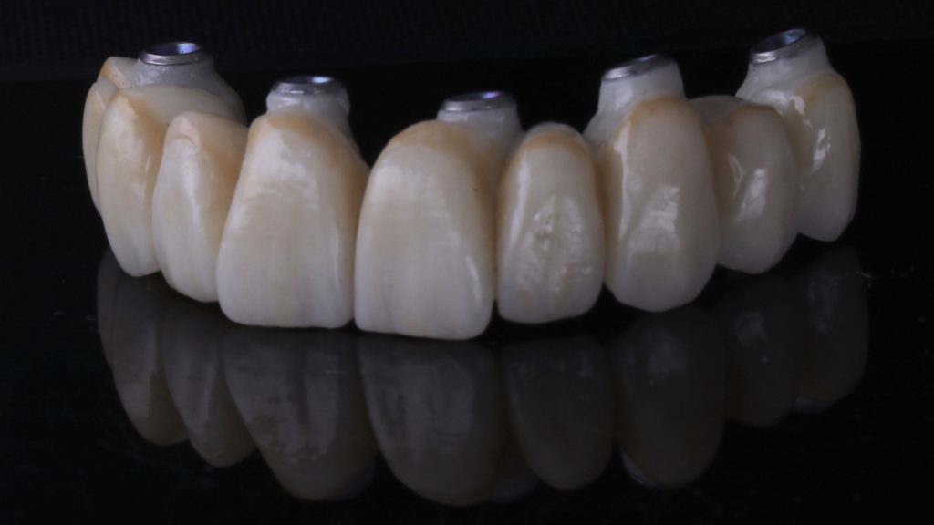
Figure 30 The final prosthesis

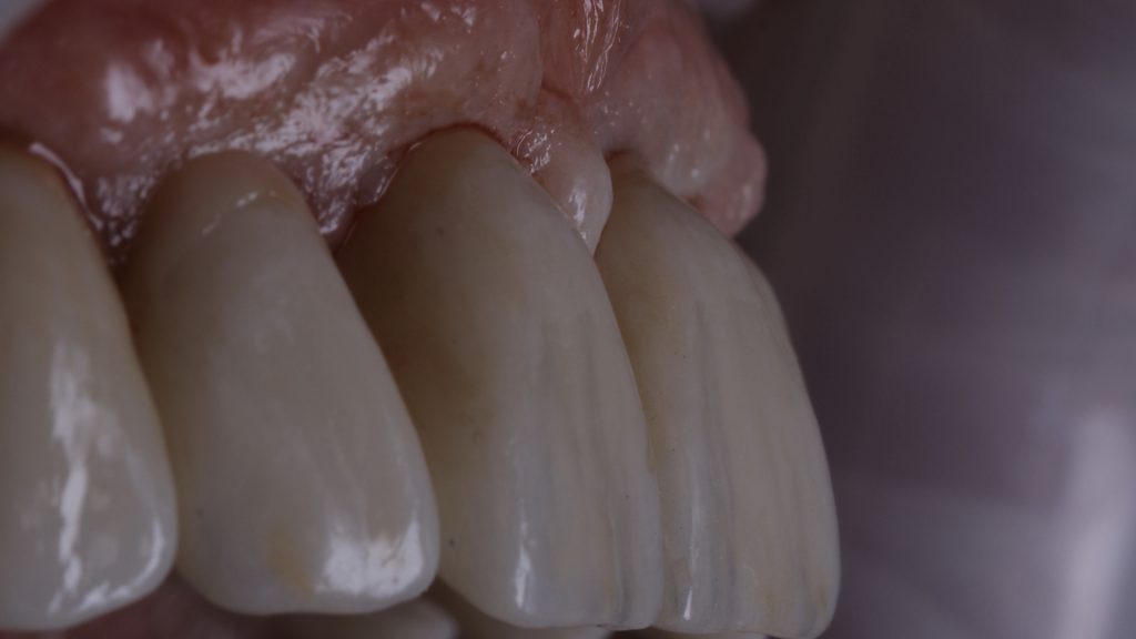
Figure 31 Soft tissue integration post-implant restoration

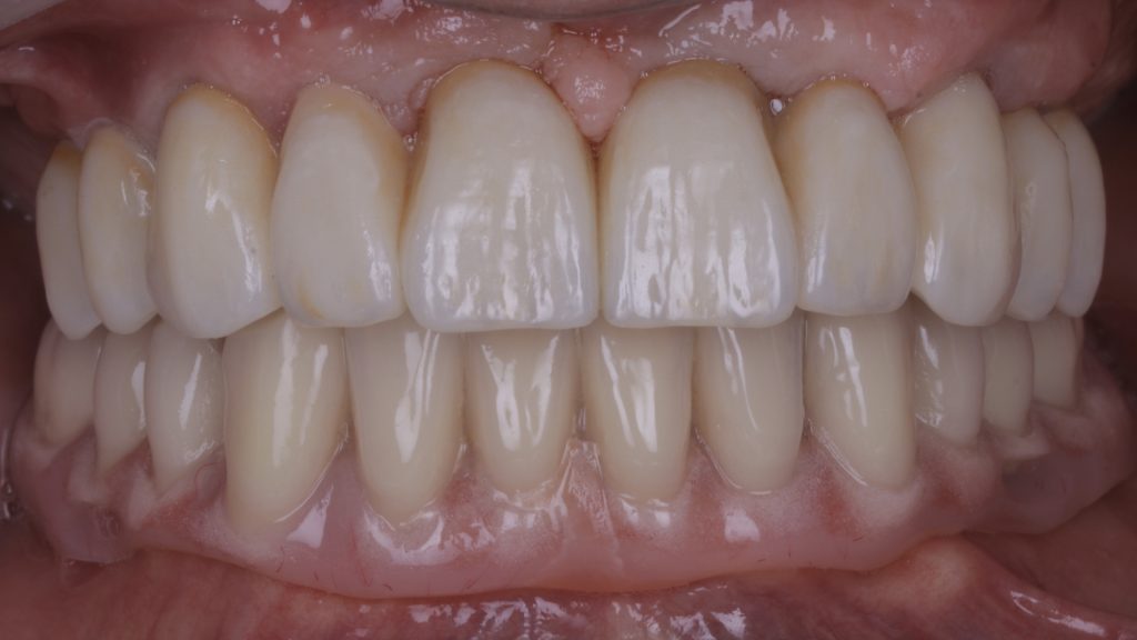
Figure 32 The final result

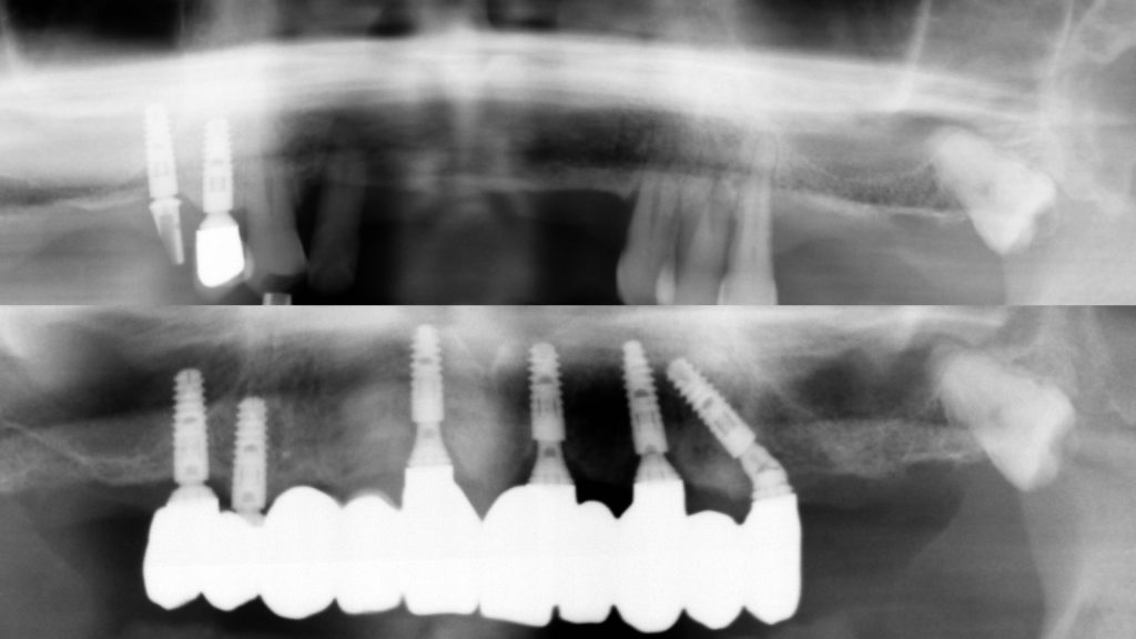
Figure 33 Pre- and post-implant restoration radiographs


Review

The patient was overjoyed with the final outcome. This case demonstrates that by following a minimally invasive approach to full-arch implant treatment, a natural-looking result can be achieved that is superior to traditional fixed dentures. Through meticulous treatment planning, soft tissue manipulation and prosthetic design, patients can benefit from a highly aesthetic and functional FP1 restoration.[ii], [iii] Practitioners can learn to carry out treatment predictably with The FP1 Course – an innovative new programme that explores a muco-gingival approach to full-arch dentistry, incorporating both digital and analogue processes. For course dates and to book your place, visit The FP1 Course Facebook page at www.facebook.com/FP1Course/


For more information visit www.tendental.com or call on 020 33932623


Author bio:

Dr Martin Wanendeya heads the award-winning Ten Dental+Facial team alongside colleague, Dr Nikhil Sisodia. Martin is a seasoned and knowledgeable implant surgeon with experience in the latest techniques.

[i] Hanser, T. and Khoury, F. (2016) Alveolar Ridge Contouring with Free Connective Tissue Graft at Implant Placement: A 5-Year Consecutive Clinical Study. The International Journal of Periodontics & Restorative Dentistry. 36(4): 465-473.

[ii] Liu, C-L. S. (2004) Use of a Modified Ovate Pontic in Areas of Ridge Defects: A Report of Two Cases. J Esthet Restor Dent. 16: 273-283. 

[iii] Pozzi, A., Tallarico, M. and Moy, P. K. (2015) The Implant Biologic Pontic Designed Interface: Description of the Technique and Cone-Beam Computed Tomography Evaluation. Clinical Implant Dentistry and Related Research. 17(2): e711-e720.


A question of sport

As the 2020 Summer Olympics in Tokyo draw closer, the debate on how sport can have an impact on oral health is once again thrown into the fray. This has always been a hotbed of discussion, but now there is new evidence to suggest that the situation may perhaps be worse than was originally anticipated.[i]

The research carried out by the UCL Eastman Dental Institute found that of 352 Olympic and professional athletes surveyed, 49.1% had untreated tooth decay, while 32% felt that their oral health was having a negative impact on training and performance. A large majority also had early signs of gum inflammation,i but what’s most worrying of all is that this is in spite of good oral health practises.

As with other aspects of their life, it turns out that professional athletes are, for the most part, fairly fastidious about oral hygiene, with the report stating that 94% brushed their teeth at least twice daily and 44% regularly cleaned interdentally. When you consider that the figures for the general population are at 73% for twice-daily brushing and 27% for interdental cleaning,[ii] it’s amazing to think that athletes’ oral health is in such peril.

Of course, the problem isn’t necessarily because of their attitude towards oral health and daily routine, but rather as a result of practises associated with training and competing – diet being the main culprit. In order to compete at a high level, athletes are required to follow a strict regime that includes, amongst other things, sufficient consumption of carbohydrates that provide energy and healthy sources of protein.[iii] Typical foods include brown rice, fruit, pasta, fish, lean meat, eggs, beans and lentils, as well as cheese, yoghurt and milk – all of which are considered healthy when consumed as part of a balanced diet. However, during training and competitions, athletes also tend to consume snacks such as protein bars. While these are convenient to eat on the go and helpful in maintaining energy levels, they can be harmful to oral health – the reason for this being that they contain so much sugar. In fact, the sugar content of an average protein bar could rival that of some of the most notorious sugary treats.[iv]

The same goes for energy gels and sports drinks, which also form part of most athletes’ diets.i Between the sugar content and acidity (one study showed that this can range from 9.74-13.44 mls of 0.1M NaOH) of these drinks,[v] athletes are at extremely high risk of dental caries and tooth erosion – not to mention that the proinflammatory effects of a high carbohydrate diet can also increase the chance of periodontal disease.[vi] However, it’s not just diet that athletes have to be mindful of when it comes to their oral health – at least in certain sports anyway.

For athletes where there is a lot of airflow involved with their sport, it is not uncommon for saliva flow to be reduced, leading to local drying of the mouth and, in turn, complications such as caries and oral infections.[vii] This means that for cyclists, runners, triathletes and the like, there’s a lot more to worry about than just what they’re consuming. Let’s not forget either how athletes such as footballers have been found to be suffering in recent years. It was only in 2015 that a study revealed nearly 4 out of 10 top-level UK footballers surveyed had advanced tooth decay and 1 in 20 had irreversible periodontal disease.[viii] With all this in mind, it is clear that greater action and intervention is needed if athletes’ oral health is to be improved moving forward.

The news that an intervention study has already been piloted then – and that dental professionals have provided advice on increased fluoride use, more frequent dental visits and replacement of energy supplements with more oral-friendly alternatives – will be music to many people’s ears. What remains is for these recommendations to be passed down to amateur athletes so that they too can protect themselves against potential risks and maintain good oral health. While a typical patient may not be at risk on the same scale as a professional athlete or Olympian would, there is a chance that their oral health and daily regime could be in a worse state to begin with. We know, after all, that professional athletes typically have better oral hygiene routines than the general public.ii

 If this is the case with your patients, or you are currently treating people who you know are active participants in sports, then an explanation of effective practises and demonstration of the correct techniques will go a long way. To achieve an effective clean easily and safely, Curaprox recommends the Hydrosonic Pro electric toothbrush. Boasting gentle CUREN® bristles and an intuitively designed brush head that enables the user to clean hard-to-reach areas, the Hydrosonic Pro is an ideal choice for any patient that needs a helping hand in maintaining their oral health.

Together with the profession’s continued vigilance on monitoring the nation’s oral health, from professional athletes and Olympians to the wider community, let us hope that we see some improvement in 2020 and beyond.


For more information please call 01480 862084, email info@curaprox.co.uk or visit www.curaprox.co.uk


[i] Julie Gallagher, Paul Ashley, Aviva Petrie & Ian Needleman. Oral health-related behaviours reported by elite and professional athletes. British Dental Journal, 2019. Accessed online 16th October 2019 at https://www.nature.com/articles/s41415-019-0617-8

[ii] Simply Health. Consumer Oral Health Survey 2019. Accessed online 16th October 2019 at https://www.simplyhealth.co.uk/content/dam/simplyhealth/denplan/documents/simplyhealth-COHS-2019.pdf

[iii] NHS. Food and drinks for sport. Accessed online 10th October at https://www.nhs.uk/live-well/eat-well/food-and-drinks-for-sport/

[iv] The Fitness Food Index. “Are protein bars really healthy?”. Accessed online 16th October 2019 at https://www.protectivity.com/fitness-food-index/

[v] Rees J, Loyn T, McAndrew R. The acidic and erosive potential of five sports drinks. Eur J Prosthodont Restor Dent. 2005;13(4):186-90. Accessed online 16th October 2019 at https://www.ncbi.nlm.nih.gov/pubmed/16411577

[vi]  Chapple ILC. Potential mechanisms underpinning the nutritional modulation of periodontal inflammation. J Am Dent Assoc 2009;140:178–84. Accessed online 16th October 2019 at https://www.ncbi.nlm.nih.gov/pubmed/19188414

[vii] Dodds M, Roland S, Edgar M, Thornhill M. “Saliva: A review of its role in maintaining oral health and preventing dental disease. BDJ Team volume 2, Article number: 15123 (2015). Accessed online 16th October 2019 at https://www.nature.com/articles/bdjteam2015123

[viii] Needleman I, Ashley P, Meehan L, Petrie A, Weller R, Mcnally S, Ayer C, Hanna R, Hunt I, Kell S, Ridgewell P, Taylor R. Poor oral health including active caries in 187 UK professional male football players: clinical dental examination performed by dentists. Br J Sports Med. (2015). Accessed online 16th October 2019 at bjsm.bmj.com/content/early/2015/10/01/bjsports-2015-094953.short?g=w_bjsm_ahead_tab

Effortless treatment positioning

Clark Dental offers an intelligently designed dental unit from Sirona that provides the option of changing the treatment position quickly and effortlessly.

The Intego Ambidextrous can be switched from a right- to a left-handed treatment position in just 15 seconds, without requiring any additional assembly or space in surgery. This is possible thanks to the dental unit’s compact design, which makes it ideal for multi-clinician practices.

With its advanced ErgoMotion feature, the Intego Ambidextrous ensures patients are fully supported for a more comfortable treatment experience. You also benefit from the most ideal working position, optimal visibility and integrated equipment options for a superior operating environment.


For more information call Clark Dental on 01268 733 146, email info@clarkdental.co.uk or visit www.clarkdental.co.uk

Common infections and oral health

Common sexually transmitted infections (STI) have health consequences within the oral cavity and beyond. These infections can be transferred non-sexually, which underscores the need for thorough decontamination and sterilization protocols.

Chlamydia

Chlamydia is the most common bacterial STI in existence. When present in the pharynx, it can cause pharyngitis or lymphadenitis, but in the majority of cases it is asymptomatic (only 14% of men experience symptoms).[1] Chlamydia trachomatis has been recorded within the epithelial lining of periodontal pockets and within the gingival sulcus of patients with periodontitis.[2]

Research has found that C. trachomatis can occupy other biofilms, such as those produced by Candida albicans (a fungal yeast which can cause infections of the mouth, throat and esophagus).[3] Biofilms containing these bacteria can develop in the oral cavity, and on inorganic surfaces such as medical devices. Biofilms can harbour C. trachomatis, allowing it to remain infectious for up to 72 hours following exposure, and serve as a reservoir for the pathogen, leading to reinfection. C. trachomatis is capable of replicating within epithelial cells, and can infect them following release from biofilm.[4]

HPV

The human papilloma virus (HPV) is the most common sexually transmitted virus, though it can be spread in other ways, including on improperly sterilized surgical instruments and other objects.[5] Approximately 20% of oral cancers and 60-80% of oropharyngeal cancer cases are believed to be attributable to HPV infection.[6],[7] HPV is well established as the leading cause of cervical cancer, which has lead to widespread immunisation of young women in the UK against the most carcinogenic strains. This programme has recently been expanded to cover men and teenage boys. However, because the programme, in some cases, requires men to request inoculation at a GUM clinic, coverage will almost certainly be less thorough than the programme for girls which occurs during compulsory education.[8]  This is changing as similar inoculation programmes are being set up in secondary schools.

Preventing cross-contamination

Following disinfection and sterilization protocols carefully and thoroughly prevents the spread of harmful microbial agents between patients, and protects your staff in the instance of sharps injuries.

With integrated traceability, the new Lisa vacuum B sterilizer from W&H can be used to handle your practice’s sterilization needs reliably and with great efficiency. W&H can also provide ECPD training, helping clinicians obtain a thorough understanding of the theory and practice of handpiece maintenance and decontamination, and ensuring they are confident in reprocessing handpieces in accordance with HTM 01-05.

The oral cavity is never going to be a perfectly sterile environment, and every mouth harbours countless species of bacteria, fungi and viruses. While patients are not unlikely to become infected with diseases such as chlamydia and HPV at some point during their lives, minimising the risk of contamination between patients is a critical duty of care and can help prevent both major and minor health repercussions.


To find out more visit www.wh.com/en_uk, call 01727 874990 or email office.uk@wh.com


[1] Chan P., Robinette A., Montgomery M., Almonte A., Cu-Uvin S., Lonks J., Chapin K., Kojic E., Hardy E. Extragenital infections caused by Chlamydia trachomatis and Neisseria gonorrhoeae: a review of the literature.  Infectious Diseases in Obstetrics and Gynecology. 2016; 5758387. https://www.hindawi.com/journals/idog/2016/5758387/ April 4, 2019.

[2] Reed S., Lopatin D., Foxman B., Burt B. Oral chlamydia trachomatis in patients with established periodontitis. Clinical Oral Investigations. 2000; 4(4): 226-232. https://www.ncbi.nlm.nih.gov/pmc/articles/PMC2760468/ April 4, 2019.

[3] Filardo S., Di Pietro M., Tranquilli G., Sessa R. Biofilm in gential ecosystem: a potential risk factor for chlamydia trachomatis infection. Canadian Journal of Infectious Diseases and Medical Microbiology. 2019; 1672109. https://www.hindawi.com/journals/cjidmm/2019/1672109/ April 4, 2019.

[4] Filardo S., Di Pietro M., Tranquilli G., Sessa R. Biofilm in gential ecosystem: a potential risk factor for chlamydia trachomatis infection. Canadian Journal of Infectious Diseases and Medical Microbiology. 2019; 1672109. https://www.hindawi.com/journals/cjidmm/2019/1672109/ April 4, 2019.

[5] Sabeena S., Bhat P., Kamath V., Arunkumar G. Possible non-sexual modes of transmission of human papilloma virus. Journal of Obstetrics and Gynaecology Research. 2017; 43(3).  https://obgyn.onlinelibrary.wiley.com/doi/full/10.1111/jog.13248 April 4, 2019.

[6] Kim S. Human papilloma virus in oral cancer. Journal of The Korean Association of Oral and Maxillofacial Surgeons. 2016; 42(6): 327-336. https://www.ncbi.nlm.nih.gov/pmc/articles/PMC5206237/ April 4, 2019.

[7] Poelman M., Brand H., Forouzanfar T., Daley E., Jager D. Prevention of HPV-related oral cancer by dentists: assessing the opinion of Dutch dental students. Journal of Cancer Education. 2018; 33(6): 1347-1354. https://www.ncbi.nlm.nih.gov/pmc/articles/PMC6280774/ April 4, 2019.

[8] NHS. Who should have the HPV vaccine? NHS. 2017. https://www.nhs.uk/conditions/vaccinations/who-should-have-hpv-cervical-cancer-cervarix-gardasil-vaccine/ April 4, 2019.