The advantages of an implant with a zirconia collar – Dr Massimo Palermo


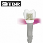
Dr Massimo Palermo explains through a case study how a tissue-level implant with a zirconia collar can help clinicians regenerate and preserve bone, while also encourage effective healing of the gingiva for superior soft tissue management.


In May 2013, a 46-year-old female patient presented with an infection (Figure 1) near the apex of the LR6 tooth root. A treatment plan was formulated and discussed with the patient, which involved extraction of the LR6 and placement of a 4mm diameter by 10.5mm length TBR Z1 dental implant.


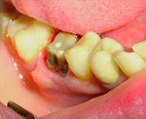
Figure 1:  Infected tooth


A radiograph (Figure 2) was taken which showed that the patient had insufficient bone quality and quantity, as the top of the cortical bone was not level. In order to preserve the socket, extraction of the LR6 was performed and a collagen sponge made of lyophilized non-denatured type-I collagen extracted fromequine Achilles’ tendon (Condress) was placed, in order to encourage accelerated healing[i]of the supporting bone so that an implant could be placed.


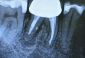
Figure 2:  Radiograph of the infected LR6


Implant placement

Five months following extraction, a radiograph (Figure 3) taken in preparation for implant placement demonstrated imperfect bone levels, despite the socket preservation procedure. The decision was therefore made to perform guided bone regeneration (GBR) at the time of implant placement.


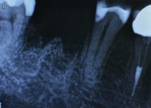
Figure 3: Post-extraction and socket preservation


The implant surgery involved making an incision in the gingiva to raise a full thickness flap, which provided access to the bone. A hole was drilled in the surgical site before the implant was placed at the pre-determined depth and diameter. At this time, a synthetic bone graft made from B-TCP (Graftek) was carried out and a type-I equine collagen membrane was placed to prevent the grafted site being colonized by soft tissue.[ii]A periosteal-release incision was created to obtain a tension-free closure over the cover screw of the implant.[iii],[iv]

Having later achieved excellent bone and soft tissue regeneration, a second surgical step was required. A circular opening was created with a diode laser so that the healing abutment (Figure 4) could be placed. While this second surgical step is not normally indicated when using the Z1 implant – the gingiva usually grows around the transgingival zirconia collar naturally[v]– it was selected in this case in order to preserve the maximum amount of soft and hard tissue.


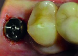
Figure 4: Healing abutment placed


Restoration

Six months post-implant surgery, a radiograph (Figure 5) demonstrated excellent bone growth that fully supported the implant. Figure 6 shows that the implant had healed well with no sign of infection or gingival inflammation. The Z1 implant facilitated enhanced healing of the gingiva, particularly around the zirconia collar.


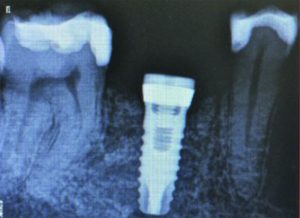
Figure 5: Radiograph of implant placed


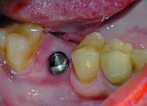
Figure 6: Six months following placement of implant


In fact, healing had been so successful that the gingiva had begun to grow over the healing cap, so a laser was used to cut back the soft tissue to improve access to the implant component. Unlike traditional bone-level implant solutions that often require two-stage surgery in order to place the restoration (as a result of the gingiva healing over the implant), the Z1 implant usually only requires one-stage surgery. This makes it more comfortable for the patient and more time-efficient for the practitioner. However, this case was an exception because the full thickness flap and the GBR procedure carried out during implant placement – combined with the outstanding soft tissue integration properties of the zirconia collar – led to excessive gingival proliferation which required laser gum surgery.

The healing cap was then removed and figure 7 shows that the white collar of the implant could be seen. If a traditional titanium implant solution is used, the greyish hue of the metal component can become visible in the case of thin gum or gingival recession. The A2 shade of the Z1 implant’s collar ensures natural transparency.


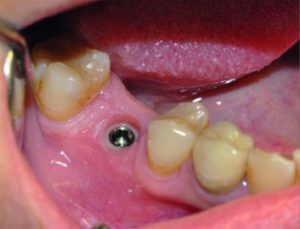
Figure 7: Healing abutment removed


Implant restoration

After two weeks, the soft tissue had healed from the laser treatment and several months after implant placement, the gingiva remained pink and healthy.

The healing cap was removed in order to fit a TBR Swiss clip direct impression coping (Figure 8). This does not involve the use of a screw and is a simple, quick impression-taking solution that combines the benefits of both direct and indirect techniques. In particular, it is ideal for closed-tray methods involving single-unit implants, meaning clinicians do not have to rely on a customised tray. An impression (Figure 9) was then taken, which was sent to the laboratory technician to create a model (Figures 10 and 11). The technician fabricated a zirconia crown (Figures 12 and 13), which was sent back to the practice for a try-in with the patient to ensure a satisfactory fit (Figures 14, 15, 16, 17 and 18).


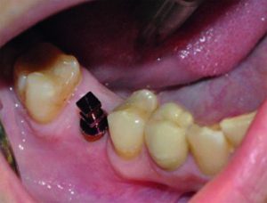
Figure 8: TBR Swiss clip impression coping


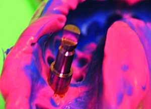
Figure 9: Impression material with impression coping


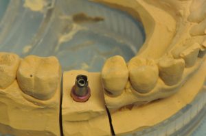
Figure 10: Abutment on impression model


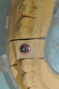
Figure 11: Overview of abutment on impression model


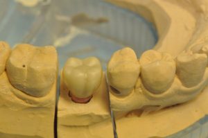
Figure 12: Zirconia crown on impression model


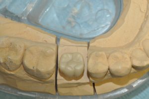
Figure 13: Overview of zirconia crown on impression model


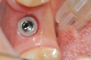
Figure 14: Implant prior to fitting the crown


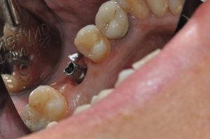
Figure 15: Abutment placed


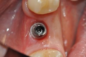
Figure 16: Overview of abutment placed


Figure 17: Crown fitted to the abutment


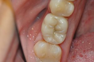
Figure 18:  Overview of the final restoration


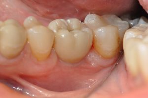
Figure 19: Comparison of the crown to the adjacent natural teeth


Once this was confirmed, temporary cement (ImplaCem Automix Precision) was used to secure the prosthesis to the abutment. Although this material is very strong and demonstrates long-term stability comparable to permanent cement, it enables easy removal of the crown if necessary.

Traditional abutments are usually the same diameter as the implant component. With regard to the Z1 implant, the abutment is always smaller than the zirconia collar, so that masticatory forces can be absorbed by the abutment and the titanium core of the implant. In the case of a bone-level implant, there would be a smaller margin for the crown to rest on, corresponding to the shoulder of the abutment. However, the tissue-level Z1 implant – whose zirconia collar ensures an optimal cervical adaptation of the crown and, in our case, a ceramo-ceramic continuity between the implant collar and the crown – helps prevent any gingival infiltration. In addition, conventional implant abutments usually cover the implant components completely, while the Z1 collar pushes the gingiva laterally, acting like a gingival scaffold that further promotes optimal gingival health.

Result

Following completion of treatment, the zirconia collar implant provided results comparable to a natural tooth. As figure 19 demonstrates, the emergence profile of the implant was very good and looked like the adjacent natural gingiva. Two radiographs were taken – one in 2018 (Figure 20) and the other in 2019 (Figure 21). Both of these showed the same result five and six years respectively following implant placement. In fact, the peak of the bone near the collar is in the same condition as when the implant was first placed. There is excellent gingival integration (Figure 22), with no bone resorption near the zirconia collar. An increase in keratinised gingiva can be seen just below the coronal margin at the level of the implant’s zirconia collar. Evidently, the patient had very good oral hygiene to help maintain a healthy implant, but this is not the only reason for lack of bone resorption and gingival recession. The design of the Z1 implant helped to ensure there was no infiltration of bacterial pathogens, thereby reducing the risk of inflammation and peri-implantitis, which is one of the main causes of implant failure.[vi]The Z1 implant not only enables clinicians to achieve effective osseointegration, but also periointegration.  


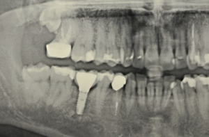
Figure 20: Panoramic radiograph of the implant and crown


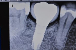
Figure 21:  Radiograph taken 6 years post-implant placement


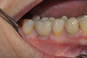
Figure 22:  The result 6 years post-implant placement


Conclusion

In the case of a titanium bone-level implant, there is often slight bone resorption and, therefore, soft tissue recession.[vii],[viii]This was not an issue in regards to the TBR Z1 implant, which performed beyond initial expectations. As a ‘soft-tissue friendly’ solution, it encourages gingival growth for effective maintenance over time[ix], as was evident in the case. It took a whole year to complete treatment because it was difficult for the patient to attend appointments at the preferred time. However, the process concluded in June 2014 and the patient is still very happy with the final result, which remains highly aesthetic and functional. 


For more information on the Z1 implant, visit tbr.dental, email support@denkauk.com or call 0800 707 6212


Author bio:

Dr Massimo Palermo, MDDS, has been working alongside TBR for over 25 years. He specialised in periodontal regeneration and reconstructive plastic surgery at Florence University. He practises from a private clinic in Rome, focusing on oral rehabilitation surgery, guided bone regeneration (GBR), guided tissue regeneration (GTR), and dental implantology. At the Sapienza University of Rome, he collaborates on research projects with the Department of Dentistry and Maxillofacial Sciences. He also works with the Faculty of Economics and Commerce, as well as the Faculty of Medicine and Surgery, contributing to research on bioethics and management in health economics.

Beyond this, Dr Palermo participates in both national and international dental courses. He trains other dentists in implant surgery, as well as GBR and GTR techniques, having authored and co-authored numerous articles on the same topics. Dr Palermo also lectures on a wide range of subjects, including oral surgery, periodontal surgery, implant surgery, regenerative bone surgery, unconventional medicine, bioethics, dental marketing and management.


References

[i]Beghé, F., Menicagli, C., Neggiani, P., Zampieri, A., Trallori, L., Teta, E., Rosini, S. Lyophilized non-denatured type-I collagen (Condress) extracted from bovine Achilles’ tendon and suitable for clinical use. Int J Tissue React. 1992;14 Suppl:11-9. [Last accessed: 20.12.18]

[ii]Elgali I, Omar O, Dahlin C, Thomsen P. Guided bone regeneration: materials and biological mechanisms revisited. Eur J Oral Sci. 2017;125(5):315-337. [Last accessed: 20.10.18].

[iii]Romanos GE. Periosteal releasing incision for successful coverage of augmented sites. A technical note. J Oral Implantol. 2010;36(1):25-30. doi: 10.1563/AAID-JOI-D-09-00068. [Last accessed: 20.12.18]

[iv]Steigmann M, Salama M, Lau K. (2012) Periosteal pocket flap for horizontal bone regeneration: A case series. Int J Perio Res Dent. 32(3):311-20 https://www.researchgate.net/publication/221694653_Periosteal_Pocket_Flap_for_Horizontal_Bone_Regeneration_A_Case_Series[Last accessed: 20.12.18]

[v]Bianchi, A. E., Bosetti, M., Doci, G., Sberna, M. T., Sanfilippo, F. and Cannas, M. (2004) The Zirconia solution: increasing osteoblasts and fibroblasts adhesion and proliferation 

[vi]Prathapachandran, J. and Suresh, N. (2012) Management of peri-implantitis. Dental Research Journal. 9(5): 516-521. Link: https://www.ncbi.nlm.nih.gov/pmc/articles/PMC3612185/. [Last accessed: 01.10.18].

[vii]Vouros, I. D., Kalpidis, D. R., Horvath, A., Petrie, A. and Donos, N. (2012) Systematic Assessment of Clinical Outcomes in Bone-Level and Tissue-Level Endosseous Dental Implants. J Oral Maxillofac Implants. 27: 1359-1374. Link: https://pdfs.semanticscholar.org/013a/5b3d054d07a78a5919fdca0df923a79b66af.pdf. [Last accessed: 01.10.18].

[viii]Le, B., Borzabadi-Farahani, A. and Nielsen, B. (2016) Treatments of Labial Soft Tissue Recession Around Dental Implants in the Esthetic Zone Using Guided Bone Regeneration With Mineralised Allograft: A Retrospective Clinical Case Series. Journal of Oral and Maxillofacial Surgery. 74(8): 1552-1561. Link: https://doi.org/10.1016/j.joms.2016.04.015. [Last accessed: 01.10.18]. 

[ix]Bianchi, A. E., Bosetti, M., Dolci, G. Jr., Sberna, M. T., Sanfilippo, S. and Cannas, M. (2004) In vitro and in vivo follow-up of titanium transmucosal implants with a zirconia collar. J Appl Biomater Biomech. 2(3): 143-150.

R A Medical Services recognised in the Parliamentary Review 

R A Medical started in 1994 as a direct heritage of Cyprane, Keighley; our Company being formed 25 years ago and now occupying a market leader position across the UK & Ireland for this very specialised area of medical equipment. 

Janet Pickles, Chairwoman of R A Medical Services Ltd., reports… 

Inclusion in this year’s Parliamentary Review is a recognition of all the hard work and subsequent achievements.  We are looking forward to attending the Houses of Parliament in October for a Gala Evening in late October.

Each year, The Parliamentary Review aims to showcase the best of business, journalism and politics, making the September release an eagerly anticipated exercise when it comes to demonstrating platforms of success and innovation across British industry. The Parliamentary Review has been an opportunity for the best and brightest business leaders, educators and healthcare professionals to speak directly to their peers and the political classes.

Success for businesses of any size does not always come easily, and this year’s edition of The Parliamentary Review is indispensable for anyone who seeks to make a name for themselves in industry. It highlights significant developments and concerns for business leaders up and down the country.

The co-chairman of The Parliamentary Review, Lord Pickles, has praised the upcoming Review as one of the most comprehensive yet. He commented that as Britain undergoes changes, it is “essential that politicians have a firm understanding of the challenges with which British organisations must contend” and that The Review once again provides a perfect platform for this.

Writing in The Review, The Chancellor of the Duchy of Lancaster, Michael Gove says “this year’s Parliamentary review comes at a momentous time for parliament, as we collectively determine the destiny of the United Kingdom.”

The best practice article for R A Medical Services can be viewed here:

https://www.theparliamentaryreview.co.uk/organisations/r-a-medical-services

ALIGN TECHNOLOGY ANNOUNCES ‘DIGITAL EXCELLENCE SERIES – POWERED BY ITERO’ FOR DENTAL PROFESSIONALS ACROSS THE UK

Align Technology, Inc. (NASDAQ: ALGN) announced today that throughout October 2019 it will be hosting the Digital Excellence Series – Powered by iTero, a new event series for dental professionals in the United Kingdom. The events will be hosted in Birmingham, Motherwell, Manchester and London, with a focus on the benefits of the iTero intraoral scanner and dental professionals’ most frequently asked questions regarding how to transform their practice from a traditional, analogue model, towards a digital one. Speakers from across the UK will share details about their journey from analogue to digital, key learnings, and the difference it has made to them, their patients, and their practice.

Key topics will include:

  • Why embrace digital technology – what are the benefits for my practice?
  • The advantages of digital workflows versus analogue workflows
  • Understand the full capabilities of iTero, and how an iTero digital scanner brings value across the entire digital workflow
  • The benefits and impact of a digital workflow on the patient journey
  • The benefits of the latest iTero innovation, iTero Element 5D with NIRI technology

The dates and locations for the Digital Excellence Series of meetings are as follows:


  • October 12th, Birmingham (09:30 – 12:30)

Venue: Align Technology UK Office

Speakers: Dr. Matt Perkins & Ashley Byrne


  • October 16th Motherwell, Scotland (18:30 – 21:00)

Venue: Dakota Hotel Eurocentral

Speakers: Dr. Elaine Halley & Steve Campbell


  • October 23rd, Manchester (18:30 – 21:00)

Venue: Victoria Warehouse

Speakers: Dr. Marcos White & Steve Campbell


  • October 30th, London (18:30 – 21:00)

Venue: Rocket Space

Speakers: Dr. Eoin O’Sullivan & Dr. Mohsen Tehranian


The events are complimentary to attend; however, registration is required as spaces will be limited.

To register or discuss these events in more detail, please contact your local Invisalign or iTero representative, or Stevie Broomfield: sbroomfield@aligntech.com  07500993440.

“HyFlex™ EDM files give me peace of mind”

Mike Dodd has undertaken posts in both general and restorative dentistry. He was also

appointed Clinical Lecturer in Restorative Dentistry at Liverpool University Dental Hospital in 2013 and then Speciality Registrar in Endodontics in 2014. Now focussing entirely on private practice, Mike is part of the team at Rose Lane Dental Practice, Liverpool, taking referrals on all aspects of endodontics. He says:

““The Hyflex™EDM file system is flexible – literally and figuratively. I can complete most cases with just one or two files, from routine, everyday endodontic cases to cases with curved, narrow or calcified canals. The system covers all the bases; the files can be pre-bent when access is difficult and pre-curved to bypass ledges. It’s versatile too, with options available for glidepath management and large apical sizes.  

“Hyflex™EDM files are robust, with a hard outer coating and tough inner core for peace of mind. My mainstay is the HyFlex™ EDM 25/~ OneFile. For cases with curved canals, I find the addition of the 20/.05 fileand the 10/.05 Glidepath file to be particularly useful.

“Compared to other files on the market, the Hyflex™EDM system is cost effective, too.”


To find out more visit www.coltene.com,

email info.uk@coltene.comor call 01444 235486

Get organised, get motivated, get informed and get ahead!

The Carestream Dental Summit 2019 will feature a wealth of fresh ideas and product breakthroughs for the whole team to benefit from.

Inspirational sessions will be delivered by leading professionals in the field, including:

Martin Attariani

Jan Einfeldt

Jaswinder Gill

Sana Luqmani

Gulshan Murgai

Adam Nulty

Emma Owen

Pawel Paskiewicz

Hugo Patrao

Paul Reaney

Eric Rooney

Kunal Shah

Andrew Taylor

Patrik Zachrisson

They will share their extensive expertise regarding digital imaging, intraoral scanning and dental practice management, helping all practice teams take their businesses to the next level.

For more information or to book, visit www.carestreamdental.com/uksummit.


For more information, contact Carestream Dental on 0800 169 9692 or

visit www.carestreamdental.co.uk

For the latest news and updates, follow us on Twitter @CarestreamDentl

and Facebook

The benefits of industry involvement with professional education – PG Cert in Implant Dentistry

There are many benefits afforded by a close working relationship between dental implant education providers and product suppliers. Perhaps the greatest advantage is that clinicians can learn how to use specific implant systems. They can discover all the science behind the products and gain practical experience in order to achieve the best possible results with each solution when in practice. In addition, product suppliers often work with some of the most respected experts in their fields. Not only does this afford confidence in the quality of systems presented during training, but it also means that world-class speakers are often available to share their extensive knowledge and experience with delegates. All of this serves to provide a higher quality of tuition and support for dentists looking to develop their skills.

For the very best learning experience, it’s important that professional training covers both theoretical and practical elements, with surgical and restorative aspects. Having a comprehensive understanding of all the procedures involved will enable dentists to more confidently provide patients with the information they need for informed consent.

This therefore requires knowledge of both analogue and digital treatment techniques. Digital is certainly the future of implantology, and indeed likely the future of all dentistry. While not every dentist will be able to introduce a completely digital workflow straight away, being aware of the options and understanding the potential benefits is still an important part of good training.


Building bridges

TRI®is an innovative Dental Implant Solutions Provider from Switzerland, with a strong commitment to professional education. We believe that only with quality training will dentists be able to maximise on the benefits provided by the best implant systems on the global market. We were therefore delighted to get the opportunity to work alongside Professor Cemal Ucer, who uses the TRI®Dental Implant System himself. Over time we have built a fantastic relationship and friendship with Professor Ucer, enabling us to work together on various courses. Among these is the PG Cert in Implant Dentistry delivered through Ucer Education.

This 12-month course provides dentists with the knowledge and practical skills they need to place and restore implants successfully. Importantly, it covers all the basics, with information and support tailored to the needs of those beginning their journey with implants. The practical stages allow delegates to physically place implants in models and under supervision, ensuring they can safely deliver treatment to their patients. For more experienced delegates, complex topics such as bone augmentation and soft tissue management are covered and on-going mentoring ensures continued support as they develop their skills.

Accredited by EduQual at Level 7, the course is presented by world-class speakers, including Professor Ucer himself. Training utilises the cutting-edge facilities of the ICE Postgraduate Dental Institute in Manchester. It highlights the advantages of a digital workflow, while still covering analogue techniques to cater for everyone. In addition, it provides an opportunity for dentists to get to grips with leading implant solutions, including the TRI®System.

Trusted products

Easy to use products that facilitate a simple workflow encourage good results by reducing the potential margin for error. That’s why the TRI®Dental Implant System was designed with simplicity in mind, offering an intelligent yet streamlined portfolio. The TRI®Performance Concept incorporates the company’s ethos to help deliver predictable osseointegration, immediate stability, a strong connection and long-term aesthetics for every patient.

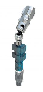
All products are of the highest standards, demonstrating the height of Swiss quality. Of note is the newly patented 3D-Touch Open-Tray Impression Post – an intra- and extra-oral scanbody that suits both analogue and digital impressioning methods for workflow flexibility. The narrow component is ideal for even small spaces and it attaches to the implant without a screwdriver for maximum ease of use. It is also easy to unscrew at any angle of up to 45 degrees, big advantage especially in the posterior region. Furthermore, TRI®wholeheartedly supports the digital workflow with its TRI®Digital Implant. The world’s first implant to be created specifically for digital manufacturing, the concept makes it easy to design the restoration directly on the implant, with no need for an abutment.

Ultimately, the innovative TRI®Dental Implant System is cost-efficient, fast, simple and intuitive – ideal for both beginners and experienced clinicians looking to excel in implantology. The supplier also supports its users with an extensive library of online tutorials, webinars and further training courses, ensuring all dentists get the very most out of the TRI®Dental Implant System.


Education at its best

If you’re interested in developing your dental implant skills with a trusted training provider and world-class products, the PG Cert in Implant Dentistry from Ucer Education could be for you. The programme is a result of careful thought and extensive experience, helping to set you up for a long and successful career in dental implantology.

For more information on the PG Cert in Implant Dentistry from Ucer Education – which is supported by several leading industry partners, including TRI®Dental Implants – please visit www.ucer.education or call 0161 237 1842


For further details about the TRI®implant portfolio, please visit www.tri.swissor email info@tri-implants.swiss


Dentistry Show All things endo and perio

The Speciality Interest Theatre at the British Dental Conference and Dentistry Show 2019 provided a wealth of information and advice in the areas of endodontics and periodontology.

Leading professionals in the field explored intriguing topics, including Mark Ide, Sanjeev Bhanderi and Peter Galgut.

Tatsiana Samalazava, dentist from Mount Wise Dental Practice, commented:

“This was my first time at the British Dental Conference and Dentistry Show and I found it very informative, covering a wide range of topics. I loved the lectures on endodontics and periodontal disease.”


The British Dental Conference and Dentistry Show, co-located with DTS.

2020 Dates: 15th– 16thMay NEC Birmingham

 For all the latest information on the next event, visit www.thedentistryshow.co.uk, call 020 7348 5270 or email dentistry@closerstillmedia.com

Dentsply Sirona on a worldwide mission for dental care

By supplying innovative equipment, Dentsply Sirona International Special Clinic Solutions (ISCS) empowers dental professional to provide better dental care all over the world – even in remote and underserved regions. 

From the Maldives to Nigeria to Malawi – in 2018, Dentsply Sirona completed successfully three exceptional clinic projects. Now, the patients in these regions can benefit from advanced dental care thanks to modern equipment. “It is important for us, to contribute to the development of dental care services in less-favored countries by providing technologies that improve access to better oral health”, explains Joerg Vogel, Vice President Sales International Clinic Solutions at Dentsply Sirona. These projects reflect Dentsply Sirona’s presence in over 40 countries worldwide and – in terms of sales presence – in more than 120 countries.  

On the Maldives, Dentsply Sirona delivered treatment centers, a 3-D imaging unit as well as CAD/CAM systems such as CEREC and inLab to the Indira Gandhi Memorial Hospital in the capital Malé. The innovative equipment will serve both, the local inhabitants and the tourists. As the largest governmental healthcare facility in the Maldives with approximately 300 beds and a total of 21 departments the clinic provides services to about 150,000 inhabitants.  

Another project is part of Dentsply Sirona’s corporate social responsibility program to improve access to dental health care in underserved areas. In the Sengaby Baptist Medical Clinic in Salima, central Malawi, Dentsply Sirona donated and installed one Intego treatment center. Established in the early 1970s, the Baptist Medical Clinic is the only facility that provides dental services in the Salima district besides the district’s hospital. The clinic provides free dental services since the average income of most of the surrounding people is less than one US dollar per day.

In the third project example, Dentsply Sirona equipped the Dental Department of the Nigerian Navy Reference Hospital Ojo /Lagos (NNRH Ojo) with high-quality dental technologies. The new dental equipment consists of Teneo and Intego Pro treatment centers as well as intraoral, 2-D and 3-D imaging units and last but not least the CAD/CAM systems CEREC and inLab. At the official inauguration ceremony, his excellency Muhammadu Buhari, President and Commander-in-Chief of the Armed Forces of the Federal Republic of Nigeria, thanked Dentsply Sirona and encouraged other states in Nigeria to take the Ojo project as a good example. So, similar dental equipment like in the Ojo example is currently on its way to the Nigerian Navy Reference Hospital in the capital Lagos where it will be implemented until the middle of the year. Besides the Navy’s employees and their relatives, the state-of-the-art dental care center will also be accessible in terms of public oral health services. Therefore, a team of the Nigerian Navy dental professional and technicians completed a comprehensive clinical and technical educational program at Dentsply Sirona’s training facilities in Bensheim, Germany.

Dentsply Sirona is the world’s largest manufacturer of professional dental products and technologies and has been in existence for more than a century.

A bone to pick – Dr Nik Sisodia

A strong foundation, metaphorically or literally, is highly influential on long-term success. This truism very much applies to dental implants, where the foundation – bone tissue – must be of sufficient quality and quantity or else they may not successfully osseointegrate enough to prove viable in the long term.[1]

Osseointegration

Osseointegration is the process by which the bone surrounding the surface of a load-bearing implant knits to it. Stability is critical to the success of this process. While primary stability is attained from the mechanical adhesion of the implant, secondary stability is contingent on primary stability, and is the result of the osseointegration process. Where primary stability is lacking, the bone remodelling process will be adversely affected, leading to more fibrous bone and a weaker implant-bone interface. Micromotion due to a lack of stability can ultimately lead to localised bone resorption.[2]

Successful osseointegration is reliant on numerous factors, including but not limited to: the health of the patient, smoking status, the design and material composition of the implant, differences in surgical method and execution, hormones, etc. Two of the major factors governing the long-term success of the osseointegration process is the quality and quantity of the bone the implant is inserted into.[3]


Bone quality

Not all bone is created equal. It varies in density, vascularity and so on. Generally speaking, primary stability increases with greater bone density, and this in turn can improve secondary stability (by facilitating the osseointegration process). The anatomical location of the bone influences its density. For example, the anterior mandibular bone is denser than the posterior maxillary bone. As we might expect, implants in the former are reportedly more successful.[4]

Some bone has more potential to regenerate, as is the case with predominantly trabecular (type III) bone. In contrast, densely compact, homogenous (type I) bone features a comparatively low rate of cell proliferation, alongside minimal collagen deposition and no mineralisation. Consequently, in type I bone, the tissues do not sufficiently stiffen, resulting in the level of strain induced by the implant remaining high. High-strain provokes the formation of fibrous tissue, which is far from ideal.[5]

Because primary stability can be significantly influenced by bone quality, some researchers recommend using imaging technologies like CT or CBCT to assess the bone during treatment selection and planning, in order to more reliably evaluate it and better factor it into the design choice of the implant.[6]

Insufficient bone density can also increase the chances of an iatrogenic injury, particularly if unexpected. This can lead to the surgeon placing too much pressure on the drill, relative to what is required – resulting in over-penetration and potentially a nerve injury. These types of errors can be better avoided through careful preoperative planning and assessment.[7]


Bone quantity

Dental implants require sufficient bone to provide structural stability, and to withstand the forces exerted upon it. Where bone quantity is insufficient, and/or the bone has defects, osteoplasty and hard or soft tissue augmentation can be necessitated. The quantity of bone required depends on the anatomical location, the design of the implant(s), and the structure of the bone.[8]Aesthetics are another consideration, as the shape of the bone can have a significant effect on the patient’s profile (generally speaking a reduced gumline is associated with an older appearance).


Osteoporosis

Osteoporosis is a relatively common condition that results in a reduction of bone quality and quantity. Furthermore, through several mechanisms, the condition reduces the osteogenic capacity of the bone – it impairs bone regeneration and healing.[9]While osteoporosis is theorised to increase the risk of dental implant failure and is associated with higher rates of implant loss across a number of studies, there is disagreement across the available literature on the subject as to whether it is a contraindication to treatment, and to the degree of risk it represents.[10]It is advisable that longer healing times are allowed for in patients with osteoporosis.[11]

It should also be noted that a small subset of patients (0.1%) receiving bisphosphonate treatment for osteoporosis develop bisphosphonate related osteonecrosis of the jaw (BONJ). For those predisposed, invasive dental treatment often acts as a catalyst, with studies finding that tooth extraction was a precipitating factor in 38-80% of cases.[12],[13]


Expertise

For clinicians that lack the confidence, expertise or facilities to treat dental implant patients with inadequate quantity and quality of bone, it is always worth referring to a trusted clinic like Ten Dental+Facial. The award-winning team is led by highly experienced implantologists, Drs Nikhil Sisodia and Martin Wanendeya, who offer a seamless referral service for both simple or complex cases.

With careful case assessment and treatment planning, dental implants are a fantastic treatment modality for many edentulous and partially edentulous patients. Many factors must be considered in order to ensure long-term success and minimise complications, not least the patient’s bone structure. It is important to have a good understanding of the health and pharmacological status of patients undergoing any surgical procedure, particularly as many patients seeking implants are elderly and, therefore, more likely to present factors that may increase the chance of complications – osteoporosis among them.


For more information visit www.tendental.comor call on 020 33932623


Reference


[1]Juodzbalys G., Kubilius M. Clinical and radiological classification of the jawbone anatomy in endosseous dental implant treatment. Journal of Oral & Maxillofacial Research. 2013; 4(2): e2. https://www.ncbi.nlm.nih.gov/pmc/articles/PMC3886111/May 17, 2019.

[2]Javed F., Ahmed H., Crespi R., Romanos G. Role of primary stability for successful osseointegration of dental implants: factors of influence and evaluation. Interventional Medicine & Applied Science. 2013; 5(4): 162-167.https://www.ncbi.nlm.nih.gov/pmc/articles/PMC3873594/

 May 17, 2019.

[3]Goutam M., Chandu G., Mishra S., Singh M., Tomar B. Factors affecting osseointegration: a literature review. Journal of Orofacial Research. 2013; 3(3): 197-201. https://www.researchgate.net/publication/269280361_Factors_Affecting_Osseointegration_A_Literature_ReviewMay 16, 2019.

[4]Ayranci F., Sivrikaya E., Omezli M. Is bone density or implant design more important in implant stress formation in patients with bruxism? Biotechnology & Biotechnological Equipment. 2017; 31(6): 1221-1225. https://www.tandfonline.com/doi/full/10.1080/13102818.2017.1376597May 16, 2019.

[5]Li J., Yin X., Huang L., Mouraret S., Brunski J., Cordova L., Salmon B., Helms J. Relationships among bone quality, implant osseointegration, and Wnt signaling. Journal of Dental Research.2017; 96(7): 822-831. https://www.ncbi.nlm.nih.gov/pmc/articles/PMC5480808/May 16, 2019.

[6]Howashi M., Tsukiyama Y., Ayukawa Y., Isoda-Akizuki K., Kihara M., Imai Y. Sogo M., Koyano K. Relationship between the CT value and cortical bone thickness at implant recipient sites and primary implant stability with comparison of different implant types. Clinical Implant Dentistry and Related Research. 2014; 18(1): 107-116. https://onlinelibrary.wiley.com/doi/abs/10.1111/cid.12261May 16, 2019.

[7]Steinberg M., Kelly P. Implant-related nerve injuries. Dental Clinics of North America. 2015; 59(2): 357-373. https://doi.org/10.1016/j.cden.2014.10.003May 17, 2019.

[8]Juodzbalys G., Kubilius M. Clinical and radiological classification of the jawbone anatomy in endosseous dental implant treatment. Journal of Oral & Maxillofacial Research. 2013; 4(2): e2. https://www.ncbi.nlm.nih.gov/pmc/articles/PMC3886111/May 17, 2019.

[9]Alghamdi H. Methods to improve osseointegration of dental implants in low quality (type-iv) bone: an overview. Journal of Functional Biomaterials. 2018; 9(7). http://dx.doi.org/10.3390/jfb9010007May 17, 2019.

[10]Giro G., Chambrone L., Goldstein A., Rodrigues J., Zenóbio E., Feres M., Figueiredo L., Cassoni A., Shibli J. Impact of osteoporosis in dental implants: a systematic review. World Journal of Orthopedics. 2015; 6(2): 311-315. https://www.ncbi.nlm.nih.gov/pmc/articles/PMC4363814/May 16, 2019.

[11]Merheb J., Temmerman A., Rasmusson L., Kübler A., Thor A., Quirynen M. Influence of skeletal and local bone density on dental implant stability in patients with osteoporosis. Clinical Implant Dentistry and Related Research. 2016; 18(2): 253-260. https://www.ncbi.nlm.nih.gov/pubmed/26864614

May 17, 2019.

[12]MacLean F., Mason R., Downie J., Watt I., Gallagher A., Gallacher S., Hinnie J. The incidence of bisphosphonate related osteonecrosis of the jaw (BONJ) in patients treated with oral bisphosphonates for osteoporosis. Endocrine Abstracts. 2017; 50: 55. https://doi.org/10.1530/endoabs.50.P055May 17, 2019.

[13]Palaska P., Cartsos V., Zavras A. Bisphoshonates and time to osteonecrosis development. The Oncologist. 2009; 14: 1154-1166. https://theoncologist.alphamedpress.org/content/14/11/1154.full.pdf+html

 May 17, 2019.

Update your PDP with DTS 2020

When updating your PDP for 2020, don’t forget to add DTS to your schedule!

There will be two days of lab dedicated education covering everything from the latest milling machines and 3D printers to new materials, implant prosthesis techniques and orthodontic appliances.

Speakers include extremely experienced and well-respected professionals from different fields, ensuring you get the most up-to-date and useful information. There will also be hours of enhanced CPD available for the whole lab team, so DTS 2020 is one event you really don’t want to miss.

Save the dates today!


DTS 2020 – Friday 15thand Saturday 16thMay – NEC in Birmingham, co-located with the British Dental Conference and Dentistry Show.

Visit www.the-dts.co.uk, call 020 7348 5270
or email dts@closerstillmedia.com