Focus your attention on digital

According to a major international study, the Internet may be changing our brains. After reviewing evidence from dozens of studies and experiments, a global team of academics have proposed that the Internet could produce acute and sustained alterations in our attentional capabilities and memory processes.[1] It has been reported that the human attention span has shortened significantly in the last 15 years.[2] We have access to a constantly evolving stream of online information, notifications and prompts which divides our attention over multiple sources. The good news is that we seem to be better at multitasking but individuals that are constantly switching between short activities online, may have decreased capabilities when it comes to maintaining concentration on a single task. Furthermore, those affected show less grey matter in the cerebral areas of the brain associated with maintaining focus.1

As we know, the Internet provides a wealth of information at our fingertips and people have become reliant on it to gather information. Nevertheless, this could be affecting our memory. It seems that people no longer allocate cognitive resources towards remembering specific information so that it can recalled when it is needed. Instead, we make use of the Internet which acts as an external information storage and retrieval system, outstripping the capabilities of other resources such as books, colleagues and friends.  Obviously, using this type of “cognitive offloading” for memory storage frees up the brain to benefit other areas or activities. However, it may affect our ability to remember facts. Moreover, some individuals fail to distinguish between their own capabilities and that of their devices, creating the illusion of “greater than actual knowledge” in a large proportion of the population.1

From a social point of view, people connect and communicate with friends and family over the Internet using email and video calling as well as social media platforms, online gaming and other virtual settings. Some may say, that this disengages us from the ‘real world’ yet we are living in a highly connected society. In fact, 87 per cent of all adults used the internet daily in 2019 and more than 8 out of 10 people accessed it “on the go”.[3] It is clear that the digital era is here to stay and technology is becoming ever more mainstream and sophisticated. Smart phones, on-demand TV, virtual assistants and cloud computing are the norm and the ‘Internet of Things’ (IoT) is set to revolutionise our lives further by connecting devices together and allowing us to quickly collate, monitor and analyse reams and reams of data. It is no surprise therefore, that we now expect constant, seamless connectivity with instant results and have no time for glitches. Indeed, our expectations have become higher and higher and surveys confirm that UK consumers are significantly more impatient now than they were five years ago.[4]

As far as dentistry is concerned, patients want fast, convenient services and solutions. The Internet means that people can gather information about dental treatments, services and products at the click of a button and they can read online reviews, refer to social media and shop around quickly to find the best options. Accordingly, dental technology has moved forward and digital equipment and software to enhance communication, streamline the workflow and reduce time and expense has been integrated into almost all areas of the industry. Dental professionals now have the ability to work with increased levels of accuracy and predictability using high performance equipment safely, efficiently and productively. For example, digital impressions are cleaner and more accurate than conventional impressions and digital x-ray machines produce high-resolution images that can be viewed instantly. Digital files can also be uploaded and shared directly with other dental professionals and the dental lab which allows for immediate feedback. Similarly, digital data and records can be stored, archived and recalled easily and efficiently.

Computer-aided design and computer-aided manufacturing (CAD/CAM) has also improved the design and fabrication of dental restorations, particularly dental protheses. Furthermore, milling operations have come into wider use and a more extensive range of dental materials is now required to fit into the digital workflow. In response to this and ever-increasing patient expectations, Solvay Dental 360® has developed Ultaire® AKP. This is a new generation, high performance polymer that has been custom-developed specifically for the manufacture of removable partial dentures (RPDs). Ultaire® AKP fits seamlessly into the digital workflow, which means that inaccuracies are reduced and production is streamlined. Also, Ultaire® AKP is lightweight but strong, biocompatible and completely metal-free so patients can benefit from RPDs with exceptional retention along with increased comfort and fit.

There is no denying that technology enhances our lives by making everyday tasks quicker and easier, services more efficient and industry more productive. In the dental sector, digital technology is enabling dental professionals to work in smarter ways than ever before achieving improved outcomes and increased patient satisfaction. As a result, it’s time to focus your attention on digital and get ready to meet the future of dentistry.


To book a Solvay Dental 360® Professional Lunch and Learn or to find more information about Ultaire® AKP and Dentivera® milling discs, please visit www.solvaydental360.com


Phillip Silver is the UK Country Manager and Consultant at Solvay Dental 360.™ He is a specialist in medical technologies and materials with over two decades of experience in both implantable and non-implantable devices. Phillip has worked in a range of clinical fields incorporating digital techniques and introducing new and novel technology into restorative dentistry, replacement and reconstructive surgery and facial plastics.


[1] Firth J. et al. The “online brain”: how the internet may be changing our cognition. World Psychiatry. 2019 Jun;18(2):119-129. https://www.ncbi.nlm.nih.gov/pmc/articles/PMC6502424/  [Accessed 7th January 2019]

[2] Bradbury N.A. et al. Attention span during lectures: 8 seconds, 10 minutes, or more? Adv Physiol Educ 40: 509–513, 2016. https://www.physiology.org/doi/pdf/10.1152/advan.00109.2016  [Accessed 7th January 2019]

[3] Office of National Statistics. Internet access – households and individuals, Great Britain: 2019. Released 12th August 2019. https://www.ons.gov.uk/peoplepopulationandcommunity/householdcharacteristics/homeinternetandsocialmediausage/bulletins/internetaccesshouseholdsandindividuals/2019 [Accessed 7th January 2019]

[4] Fetch. UK’s tech obsession has made us a ‘Fast and Furious’ nation. Research based on findings of a YouGov poll conducted between 19th -22nd May of 2,078 UK adults aged 18+. https://wearefetch.com/cms/content/media/2016/01/Instant-Gratification-research-release-FINAL.pdf [Accessed 7th January 2019]

Is Candida Auris a threat to your practice?

Next to providing a high standard of dental care, protecting patients and staff against dangerous pathogens is a top priority in dentistry. To ensure everyone’s safety is achieved, infection control is absolutely essential, as is an up to date knowledge of the potential risks that pose a threat to the dental practice setting – of which there are many. The difficulty is that a number of microorganisms are still largely misunderstood by experts in the field, which can make eradicating them tricky.

One such pathogen is the relatively newly discovered Candida auris – a type of yeast that lives on the skin and causes infections in humans. Though discovered back in 2009 in the ear canal of a Japanese patient in Tokyo Metropolitan Geriatric Hospital, there is still a lot of mystery surrounding Candida auris, particularly in regards to its origins. Indeed, even now after a decade of study, experts are unsure why the fungus seemed to appear out of nowhere in several places around the globe at a similar time or why cases occur when they do. It’s no doubt why it has been ranked as the second most urgent threat in the 2019 AR Threats Report.[i] What we do know, however, is that Candida auris is usually reported in nursing care or hospital settings where a person’s health is already compromised. Patients who have had a central venous catheter, lines or tubes entering their body or who have recently received antibiotics or antifungal medications are thought to be most at risk.[ii] Conversely, it is rarely thought to affect someone who is considered to be in good health.[iii]


How it spreads

 As for how it is spread, it is believed that person-to-person transmission could be the primary source, though surface contamination may also play a part.iii Again, further research will be required to determine more about the behaviour of Candida auris, but for now it is safe to say that the utmost care must be taken to prevent cross contamination from occurring moving forward.


Threat to the UK and to dentistry

As it stands, there is no evidence to suggest that there have been any outbreaks of Candida auris in dental practices, but that’s not to say that it won’t happen in the future. After all, when you consider that any patient who has a diagnosis of a severe underlying disease, diabetes, chronic kidney disease or immunosuppression is at increased risk of contracting the infection, it is perfectly within the realms of possibility to suggest that dental patients could be at risk.[iv]

Certainly, there are already a number of people that have been affected in the UK, with a news story from earlier on in the year claiming that Candida auris had been reported in 25 separate NHS hospitals at the time of writing. The report went on to claim that a total of 226 cases had been recorded, with eight Britons who had passed away in British hospitals found to be infected.[v] While Candida auris has not been determined as the cause of death (the patients were already seriously ill when they became infected), there can be no denying that this mysterious super fungus poses a real threat to humankind.


Prevention is key

That’s why infection control must continue to be implemented to a high standard. To help with this, Public Health England have published some guidance for laboratories, community healthcare settings, and hospital patients and visitors, outlining a number of key steps that should be taken.[vi] Unfortunately, there are no specific guidelines for dental settings but the main points are relevant to all healthcare professionals, so practices can stand to benefit. Otherwise, the Health Technical Memorandum 01-05 provides all the information that practitioners require to ensure decontamination is properly implemented – including advice on best practice – and there are a number of recent studies available to help protect against cross contamination.

There have been a number of reports, for instance, that suggest the use of chlorine-based products are effective for environmental cleaning,[vii],[viii] while those containing sodium hypochlorite and peracetic acid have been proven to be less effective against Candida auris.[ix] In light of this evidence, it may be time for some dental practices to review the decontamination products they currently use.

Comprised of Didecyldimethylammonium chloride, Benzalkonium chloride and Polyhexamethylene biguanide and proven to have a 99.9999% kill rate, the Steri-7 Xtra range from Initial Medical is an ideal option for any practice looking to protect against Candida auris and other dangerous pathogens. There are a range of products available, including a high-level surface disinfectant cleaner, biocidal wipes and ready to use spray, ensuring that all bases are covered. All products feature a unique reactive barrier technology for ultimate protection.


Be prepared

 There can be no doubt that Candida auris, like other pathogens, is a potential threat for dentistry. However, if practices stay abreast of the latest evidence and continue to implement effective infection control protocols, the risk of cross contamination should be kept at bay.


For further information please visit www.initial.co.uk/medical or Tel: 0870 850 4045


-Ends-


About Initial Medical Waste Experts

Initial Medical is an expert in healthcare waste management, providing a complete collection, disposal and recycling service for hazardous and non-hazardous waste and offensive waste produced by businesses and organisations within the UK.

The safe management of healthcare waste is vital to ensure your activities are not a risk to human health.  Initial Medical’s healthcare waste services ensure that all of your waste is stringently handled in compliance with legislation and in accordance with Safe Management of Healthcare Waste best practice guidelines, providing you with the peace of mind that you are adhering to current legislation.

For further information please visit www.initial.co.uk/medical or Tel: 0870 850 4045

Media enquiries:

For more information, please contact:

erica@ekcommunications.net

01227 265700


[i] Centers for Disease Control and Prevention. Antibiotic Resistance Threats in the United States 2019. Accessed online 27 November 2019 at https://www.cdc.gov/drugresistance/pdf/threats-report/2019-ar-threats-report-508.pdf

[ii] Centers for Disease Control and Prevention. General Information about Candida auris. Accessed online 27 November 2019 at https://www.cdc.gov/fungal/candida-auris/candida-auris-qanda.html

[iii] Public Health England. Candida auris – a guide for patients and visitors. Published August 2017. Accessed online 27 November 2019 at https://assets.publishing.service.gov.uk/government/uploads/system/uploads/attachment_data/file/637101/Candida_auris_-_a_guide_for_patients_and_visitors.pdf

[iv] Lone SA, Ahmad A. Candida auris – the growing menace to global health. Mycoses 2019; 62 (8). Accessed online 28 November 2019 at https://doi.org/10.1111/myc.12904

[v] Evening Standard. Candida auris: Eight Britons who dies in UK hospitals were infected with Japanese super-fungus. Published 29 April 2019. Accessed online 28 November 2019 at https://www.standard.co.uk/news/uk/eight-britons-who-died-in-hospital-were-infected-with-japanese-superfungus-a4128901.html

[vi] Public Health England. Candida auris within the United Kingdom: updated guidance published. Updated 11 August 2017. Accessed online 28 November 2019 at https://www.gov.uk/government/publications/candida-auris-emergence-in-england/candida-auris-within-the-united-kingdom-updated-guidance-published

[vii] Schelenz S, Hagen F, Rhodes JL, et al. First hospital outbreak of the globally emerging Candida auris in a European hospital. Antimicrob Resist Infect Control. 2016;5:35. Accessed online 28 November 2019 at https://www.ncbi.nlm.nih.gov/pubmed/27777756?dopt=Abstract

[viii] Biswal M, Rudramurthy SM, Jain N, et al. Controlling a possible outbreak of Candida auris infection: lessons learnt from multiple interventions. J Hosp Infect. 2017;97:363‐370. Accessed online 28 November 2019 at https://www.ncbi.nlm.nih.gov/pubmed/28939316?dopt=Abstract

[ix] Kean R, Sherry L, Townsend E, et al. Surface disinfection challenges for Candida auris: an in‐vitro study. J Hosp Infect. 2018;98:433‐436. Accessed online 28 November 2019 at https://www.ncbi.nlm.nih.gov/pubmed/29203448?dopt=Abstract

Smells like community spirit

Gemma Hales, a multi-site practice manager with Colosseum Dental, recently organised an open day for patients in Maidstone. She says:

“We organised an open day to reflect the community spirit within the area and educate our neighbours about oral health. We also wanted to raise awareness of our practice and build our reputation. 

“We structured the day with different activities, which included oral health talks, a drawing competition with prizes, role playing, tours of the practice and discussions about treatments on offer.

“We welcomed a total of 65 children and 25 adults, including the Deputy Mayor and previous Mayor of Maidstone. Those who attended couldn’t have been more complimentary about the event and everybody said they would recommend our practice to friends and family.

“Overall we have adopted the community spirit and we cannot wait to get started on the next project to share education and offer some great seasonal promotions at affordable rates for all our patients to benefit from!”


For more information about Colosseum Dental, please visit www.colosseumdental.co.uk

Saliva, chewing gum and oral health

Saliva plays a critical role in oral health and function. From aiding digestion and taste, washing away food debris, to protecting teeth and gums, salivary flow is an often overlooked but invaluable component in a patient’s oral health.

A multifunctional fluid

Saliva is primarily composed of water, but is rich in calcium, phosphate and nitrate, as well as a large number of physiologically active substances that we are still in the process of understanding. As well as helping to maintain the pH balance of the mouth, the presence of calcium and phosphate in saliva naturally facilitate the remineralisation of caries lesions.[1],[2]

Saliva serves a vital function as a lubricant, particularly for the soft tissues of the mouth and mucous membrane, as well as the teeth. This helps protect them from damage and critically, helps them maintain a hydrated state.1 Saliva provides a degree of antibacterial and fungal protection by inhibiting adhesion to oral surfaces, buffering and clearing. These effects are most active and efficient when saliva levels are high (usually during and just after eating), with buffering and clearing actions dropping to barely effective levels when unstimulated.[3]

Saliva also plays a role in taste. While much of an individual’s perception of flavour actually depends on the olfactory system, saliva is essential for breaking down and transporting molecules from substances placed in the mouth to the taste buds.[4] Additionally, a protein found in saliva (gustin) is believed to be necessary for the growth and development of taste buds.[5]

Dry mouth

Xerostomia (commonly known as “dry mouth”) is a symptom of salivary gland hypofunction – though either condition can occur separately. It is associated with numerous chronic conditions, medications and treatments (particularly radiotherapy of the head and neck). Persistent xerostomia increases the risk of caries, demineralisation, mucositis, ulcers, tooth sensitivity and oral candidiasis.[6] It can also further reduce quality of life for the patient by causing halitosis or coughing; as well as difficulty and discomfort when attempting to eat, speak, swallow or use dentures.[7],[8]

There are a number of treatments for xerostomia, yet their efficacy depends upon the cause of the condition, which is not always apparent. Chewing gum can help, various types of mouthwash (avoiding those containing alcohol), artificial saliva, drinking more water, ceasing consumption of caffeine and of course, treating the underlying cause if possible. These measures can help with symptoms, but will not necessarily arrest the condition. As patients with dry mouth receive less protection than those with normal range saliva levels, avoiding acidic food is advisable[9] but most imperatively, their oral hygiene must be given adequate attention.[10],[11]

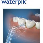
Patients with decreased salivary flow need to take particular care of their oral health. The Waterpik® Complete Care 7.0 combines a high-end Sonic Toothbrush with a state-of-the-art Water Flosser, making it perfect for patient’s daily oral hygiene routine. With both devices integrated into one convenient unit, your patients will have all the help they need to brush and floss easily and effectively.

Chewing gum and xylitol

While some people still view chewing gum as an unpleasant habit, sugar free gum is readily available and offers a considerable range of oral health benefits.

When the mouth is at rest, saliva flow drops to around 0.25-0.5 mL per minute (though this can vary quite substantially between individuals).5 Chewing gum boosts this rate more than tenfold, to approximately 5-6 mL a minute. This effect drops after the first five minutes to 2 mL/minute, then gradually reduces to 1.2-1.5 mL/minute at around the twenty-minute mark, at which level it can be sustained for the duration the gum continues to be chewed.[12],[13]

Xylitol is a naturally occurring sugar alcohol found in low amounts in various fruits and vegetables and can also be refined from birch and beech wood. It is sweeter than sugar but non-nutritive, meaning that it does not contribute to the patient’s caloric consumption. Xylitol has a number of beneficial effects on oral health. As with other sweeteners, it increases salivary flow with the benefits that confers; however, unlike other sugars and sweeteners it is for most intents and purposes ingestible by numerous strains of harmful bacteria.[14] Crucially, xylitol reduces the level of mutans streptococci found in plaque and saliva, a bacteria widely recognised as highly cariogenic.[15] Essentially, when S. mutans attempts to metabolise xylitol not only does it fail, the cell has to then expel the chemical by-product at a cost to its stored energy. This effectively starves S. mutans, leading to a substantial reduction in the levels found in the plaque and saliva of individuals who habitually chew xylitol gum, which in turn, protects against caries.14

Use of xylitol chewing gum by mothers during pregnancy and in the early months following birth has been observed to inhibit and delay transmission of S. mutans to their children, helping to protect them from caries.14

In addition to being used for certain sugar free chewing gums, xylitol is also available as a sugar substitute that can be used as a sweetener in beverages and baking. For patients with conditions that make using gum difficult or dangerous, xylitol sweets have also been successfully used instead.14 However, patients should note that its beneficial effects are diminished if combined with other sweeteners as they can fuel bacterial growth, preventing the xylitol from disrupting and starving bacteria. Furthermore, excessive consumption of sugar free gum, particularly in patients who have not built up a tolerance, can have a laxative effect.

Another useful alternative is the Waterpik® Whitening Water Flosser. As well as helping patients to floss effectively, this outstanding dental device is supplied with Waterpik® Whitening Tablets that contain xylitol to starve S. mutans and protect the teeth from caries. Whitening infuser technology mixes the tablets with water to produce a gentle whitening agent that naturally brightens the teeth by removing stains.

The take away from this is that chewing gum can help boost salivary flow and by ensuring that patients choose a gum that uses xylitol as its sweetener, these beneficial effects can be further enhanced.


For more information on Waterpik® products please visit www.waterpik.co.uk. Waterpik® products are available from Amazon, Asda, Costco UK, Boots and Superdrug online and in stores across the UK and Ireland.


[1] Kumar B., Kashyap N., Avinash A., Chevvuri R., Sagar M., Shrikant K. The composition, function and role of saliva in maintaining oral health: a review.  International Journal of Contemporary Dental and Medical Reviews. 2017; 011217: 1-6. http://www.ijcdmr.com/index.php/ijcdmr/article/view/286 January 24, 2019.

[2] Doel J., Hector M., Amirtham C., Al-Anzan L., Benjamin N., Allaker R. Protective effect of salivary nitrate and microbial nitrate reductase activity against caries. European Journal of Oral Sciences. 2004; 112(5): 424–428. https://www.ncbi.nlm.nih.gov/pubmed/15458501 January 24, 2019.

[3] Humphrey S., Williamson R. A review of saliva: normal composition, flow, and function. The Journal of Prosthetic Dentistry. 2001; 85(2): 162-169. https://www.thejpd.org/article/S0022-3913(01)54032-9/pdf January 24, 2019.

[4] Stevenson R., Oaten M., Mahmut M. The role of taste and oral somatosensation in olfactory localization.  Quarterly Journal of Experimental Psychology.  2011; 64(2): 224-240. https://journals.sagepub.com/doi/abs/10.1080/17470218.2010.491922 January 24, 2019.

[5] De Almeida P., Gregio A., Machado M., de Lima A., Azevedo A. Saliva composition and functions: a comprehensive review. The Journal of Contemporary Dental Practice. 2008; 9(3): 72-80. http://www.academia.edu/35449985/Saliva_Composition_and_Functions_A_Comprehensive_Review January 24, 2019.

[6] Plemons J., Al-Hashimi I., Marek C. Managing xerostomia and salivary gland hypofunction: executive summary of a report from the American Dental Association Council on Scientific Affairs. The Journal of the American Dental Association. 2014; 145(8): 867-873. https://www.sciencedirect.com/science/article/pii/S0002817714602002 January 24, 2019.

[7] Greenspan D. Xerostomia: diagnosis and management. CancerNetwork. 1996; 10(3). http://www.cancernetwork.com/palliative-and-supportive-care/xerostomia-diagnosis-and-management January 24, 2019.

[8] Joanna N., Thomson W. Dry mouth – an overview. Singapore Dental Journal. 2015; 36: 12-17. https://www.sciencedirect.com/science/article/pii/S0377529114200198 January 24, 2019.

[9] Joanna N., Thomson W. Dry mouth – an overview. Singapore Dental Journal. 2015; 36: 12-17. https://www.sciencedirect.com/science/article/pii/S0377529114200198 January 24, 2019.

[10] Altamimi M. Update knowledge of dry mouth- a guideline for dentists. African Health Sciences. 2014; 14(3): 736-742. https://www.ncbi.nlm.nih.gov/pmc/articles/PMC4209628/ January 24, 2019.

[11] Gil-Montoya J., Silvestre F., Barrios R., Silverstre-Rangil J. Treatment of xerostomia and hyposalivation in the elderly: a systematic review. Medicina Oral, Patología Oral y Cirugía Bucal. 2016; 21(3): e355-e366.

[12] Dodds M., Roland S., Edgar M., Thornhill M. Saliva: a review of its role in maintaining oral health and preventing dental disease. BDJ Team. 2015; 2: 15123. https://www.nature.com/articles/bdjteam2015123 January 24, 2019.

[13] Javaid M., Ahmed A., Durand R., Tran S. Saliva as a diagnostic tool for oral and systemic diseases.  Journal of Oral Biology and Craniofacial Research. 2016; 6(1): 66-75. https://www.ncbi.nlm.nih.gov/pmc/articles/PMC4756071/ January 24, 2019.

[14] Nayak P., Nayak U., Khandelwal V. The effect of xylitol on dental caries and oral flora. Clinical, Cosmetic and Investigational Dentistry. 2014; 6: 89-94. https://www.ncbi.nlm.nih.gov/pmc/articles/PMC4232036/ January 24, 2019.

[15] Banas J. Are the mutans streptococci still considered relevant to understanding the microbial etiology of dental caries? BMC Oral Health. 2018; 18: 129. https://bmcoralhealth.biomedcentral.com/articles/10.1186/s12903-018-0595-2 January 24, 2019.

Make beautiful smiles a reality

The BACD Aesthetic & Digital Dentist Theatre will be among the many educational highlights at the British Dental Conference and Dentistry Show 2020. Dr Sam Jethwa will be sharing his expertise to help delegates overcome the challenges of smile makeovers.

“My vision is to equip dentists with the skills to translate their version of a beautiful smile into reality,” he says. “My lecture will consist of creative, tried and tested solutions and protocols to satisfy the two biggest challenges that exist in smile makeover treatment: function and aesthetics.

“The British Dental Conference and Dentistry Show has an excellent line-up of lectures – especially in the BACD Aesthetic & Digital Dentist Theatre. The BACD is constantly providing the best in terms of education and this year is not one to miss.”


The British Dental Conference and Dentistry Show 2020 – Friday 15th and Saturday 16th May – Birmingham NEC, co-located with DTS.

Visit www.thedentistryshow.co.uk, call 020 7348 5270 or email dentistry@closerstillmedia.com

The BACD for young dentists

Every young dentist needs support, so have you considered joining a professional academy? 

The BACD is one of the most successful academies in dentistry. Young dentists are welcomed within an environment that believes everyone can get better at what they do, regardless of their knowledge or experience. 

As well as support, the BACD prides itself on the high-quality learning it offers its members. Cost-effective and relevant, the Academy’s educational programme will enhance your treatment delivery.

Join the BACD and you will align yourself with a network of professionals who are committed to clinical excellence. Find out more by visiting the new BACD website today.


For further enquiries about the British Academy of Cosmetic Dentistry visit www.bacd.com

Trust in our decades of experience

J&S Davis has been renowned in the dental profession for providing industry-leading products and materials for many decades. It remains as dedicated as ever to supplying solutions that will make clinicians’ lives easier.

As such, it currently offers an array of innovative product lines from some of the top manufacturers around the globe. These include the LM-Arte instrumentation designed for fine aesthetic work, the highly popular Panavia range of adhesive resins by Kuraray and the impressive Curasept ADS range of mouth rinses.

For more information on these and much, much more, don’t miss the team on stand K91 at the British Dental Conference and Dentistry Show 2020 this May!


For more information on the industry-leading products available from J&S Davis, visit www.js-davis.co.uk, call 01438 747 344 or email jsdsales@js-davis.co.uk

Lifespan and implants

Dental implants are the gold standard for rehabilitation of edentulous patients, offering the closest approximation to a brand new tooth so far devised. While dental implants enjoy a favourable success rate well in excess of 90%, there are still some cases where the implant ultimately fails.[i] Osseointegration failures are the primary cause in the short-term, while in the long-term peri-implantitis and excessive loading are the leading drivers of failure.[ii]

Nothing lasts forever and though they are durable, this is true of implants as much as anything else. Early failures due to insufficient osseointegration can be minimised through good case selection, technique and appropriate implant systems. However, dental implants are expected to serve for years in a challenging environment. While implants in more senior patients may ultimately outlast the recipient, with growing numbers of more youthful patients receiving surgery, we must consider the long-term survival of the solution.

The population is aging. Based on cohort life expectancies, it is projected that British adults today will live on average into their late 80s or early 90s (with women living around 3 years longer on average).[iii] While this projection may ultimately prove inaccurate, increasing lifespans do require us to consider not only if an implant is the best choice for a patient right now, but also how well it will serve them in the long-term. One study found implant survival rates of over 89% after 10 years, and 83% after 16 years. Following successful osseointegration most failures were attributable to peri-implantitis. Smoking was found to be a significant risk factor for implant failure.[iv]

In the long-term, compatibility may become an issue. There are already various system designs requiring a proliferation of tools. When looking two decades ahead, we cannot be certain that all current providers of these systems will remain. Opting for proven, dependable systems is the best means practitioners have of mitigating this issue. In the long-term it would be beneficial for suppliers to adopt greater standardisation.[v] As the implant may serve for well over a decade, it is not unlikely that the patient may see a different practitioner by the time problems with the implant may have developed (the patient may have moved, the dentist retired, etc.). Consequently, producing an “implant passport” could be very helpful.[vi] Such a document records details of the system used, making future clinical work easier and more efficient. For example, there are numerous types of screw in use, knowing the specific type and tool required in advance, could save valuable time.

It may be advisable to avoid permanent cementation where possible, especially in younger patients, as it can complicate future maintenance and adaptations.[vii] Dental cement has been found to lower osteoblast survival, so where required it should be used sparingly. Particular care must be taken to remove excess as its presence can harbour bacteria and provoke inflammation.[viii]

Another possible cause of failure is fracturing of the implant. This is a rare complication (around 1% of implants), but the probability increases over time. There are various factors that can lead to excessive forces acting on the implant, including screw loosening or overtightening. Parafunctional habits are a particular risk factor, with over half of patients with fractured dental implants exhibiting bruxism. Bruxism can exert high magnitude forces on the implant and for protracted periods. Ideally teeth/implants should only come into contact with each other for around half an hour per day. In patients with bruxism, contact can occur for several hours and at greater intensity. As the habit continues, the patient’s jaw muscles can strengthen from the exercise, increasing the forces at play.[ix] Bruxism can have numerous causes including sleep dysfunction, stress, psychological issues, medication and a variety of medical disorders. Parafunctional habits are a significant threat to both natural teeth and protheses and reliable treatment of causes is not always possible. One bit of good news is that the prevalence of bruxism gradually declines with age.[x]

If you are dealing with a complex case, such as a failing implant requiring surgical intervention, consider referring your patient to the Centre for Oral-Maxillofacial and Dental Implant Reconstruction. Led by Professor Cemal Ucer – Specialist Oral Surgeon – the practice offers a wide variety of advanced procedures, including nerve lateralisation and repositioning, allografts and zygomatic dental implants. With a wealth of experience and state-of-the-art facilities, your patient will receive the best of care. The clinic also provides an online advisory service and comprehensive marketing and maintenance packages for referring practitioners.

Most implant failures occur relatively early on. Provided that osseointegration is successful, implants generally survive for a considerable length of time. Being proactive in correcting early complications is therefore important to the overall success of the treatment. By ensuring that patients continue to attend regular check-ups and comply with instructions, implant failures can be further minimised. Where conditions arise that can jeopardise the implant, early detection and intervention can make a huge difference.


Please contact Professor Ucer at ice@ucer.uk or Mel Hay at mel@mdic.co

01612 371842


[i] Hickin M., Shariff J., Jennette P., Finkelstein J., Papapanou P. Incidence and determinants of dental implant failure: a review of electronic health records in a U.S. dental school.  Journal of Dental Education. 2017; 81(10): 1233-1242. https://doi.org/10.21815/JDE.017.080 November 29, 2019.

[ii] Sakka S., Baroudi K., Nassani M. Factors associated with early and late failure of dental implants. Journal of Investigative and Clinical Dentistry. 2012; 3(4): 258-261. https://onlinelibrary.wiley.com/doi/abs/10.1111/j.2041-1626.2012.00162.x November 29, 2019.

[iii] Office for National Statistics. Past and projected data from the period and cohort life tables, 2016-based, UK: 1981 to 2066. Office for National Statistics. 2017. https://www.ons.gov.uk/peoplepopulationandcommunity/birthsdeathsandmarriages/lifeexpectancies/bulletins/pastandprojecteddatafromtheperiodandcohortlifetables/latest November 22, 2019.

[iv] Simonis P., Dufour T., Tenenbaum H. Long-term implant survival and success: a 10-16-year follow-up of non-submerged dental implants. Clinical Oral Implants Research. 2010; 21(7): 772-777. https://doi.org/10.1111/j.1600-0501.2010.01912.x November 22, 2019.

[v] Sato Y., Kitagawa N., Isobe A. Implant treatment in ultra-aged society. Japanese Dental Science Review. 2018; 54: 45-51. https://doi.org/10.1016/j.jdsr.2017.12.002 November 22, 2019.

[vi] Visser A., de Baat C., Hoeksema A., Vissink A. Oral implants in dependent elderly persons: blessing or burden? Gerodontology. 2011; 28(1). https://doi.org/10.1111/j.1741-2358.2009.00314.x November 29, 2019.

[vii] Sato Y., Kitagawa N., Isobe A. Implant treatment in ultra-aged society. Japanese Dental Science Review. 2018; 54: 45-51. https://doi.org/10.1016/j.jdsr.2017.12.002 November 22, 2019.

[viii] Tatullo M., Marrelli M., Mastrangelo F., Gherlone E. Bone inflammation, bone infection and dental implants failure: histological and cytological aspects related to cement excess. Journal of Bone & Joint Infection. 2017; 2(2): 84-89. https://www.ncbi.nlm.nih.gov/pmc/articles/PMC5423581/ November 29, 2019.

[ix] Sanivarapu S., Moogla S., Kuntcham R., Kolaparthy L. Implant fractures: rare but not exceptional. Journal of Indian Society of Periodontology. 2016; 20(1): 6-11. https://www.ncbi.nlm.nih.gov/pmc/articles/PMC4795137/ November 29, 2019.

[x] Guaita M., Högl B. Current treatments of bruxism. Current Treatment Options in Neurology. 2016; 18(10). https://www.ncbi.nlm.nih.gov/pmc/articles/PMC4761372/ November 29, 2019.

The pursuit of excellence

Just fifty years ago, cosmetic dentistry was limited to invasive tooth preparation techniques that were employed to achieve the perfect Hollywood smile. Today, these unnatural-looking “tombstone teeth” are no longer the ideal, as advances in the field have extended patients’ treatment options. Dental professionals can now help patients improve the health and appearance of their smile using more conservative techniques, which often ensure that as much of the natural tooth structure as possible is preserved. With the excellent results that can be achieved from modern cosmetic dentistry, the profession can expect to see continued growth in the market as patient demand for treatment rises.

This emphasises the importance of continued learning, particularly in the face of an increasingly litigious climate. Patients are much more aware of the options available to them, but also have high demands and expectations about treatment. Dental professionals put themselves at risk of complaints and litigation unless they can demonstrate infallible record-keeping and obtaining proper patient consent. Furthermore, clinicians must show that they have the expertise to deliver consistently high standards of treatment. Therefore, the need to evidence a pathway of professional development has never been more pressing.

Continually developing skills and knowledge is especially important considering cosmetic dentistry can be challenging, particularly if patients present with multiple problems. There is no small amount of expertise and artistry involved with treatment. Having an in-depth understanding of the latest – and sometimes complex – procedures, as well as how to use materials and technology in the most effective way, is vital in order to achieve an excellent, predictable outcome.

Clinicians have to optimise dental aesthetics and function, but must also ensure that the smile is balanced with the overall appearance of the face. The modern dentist should understand and employ the most minimally invasive, long-lasting techniques to deliver the best results. This constitutes the need for practitioners to expand on their skills and knowledge within various dental disciplines, which may include restorative dentistry, prosthodontics, oral surgery and orthodontics.

Not only a leader in providing outstanding educational opportunities that support structured learning, the BACD also provides a training pathway for cosmetic dentistry through its own Wheel of Knowledge. This is designed to be a useful guide that highlights core development criteria for advancement as a cosmetic dentist. In addition, the BACD offers a chance for clinicians to achieve industry-recognised accreditation within the field. Regarded as one of the profession’s most prestigious and respected accolades, BACD Accreditation distinguishes successful candidates for their clinical excellence and improves the credibility of their cosmetic dental work among both patients and peers.

BACD members themselves are expected to ensure they continually update their skills and decision-making in the interests of patients and the Academy. That’s why they benefit from unlimited access to social and educational events such as Recommended Meetings and the celebrated Annual Conference. These events promote shared learning for the betterment of the profession overall, which ultimately embodies the BACD’s values of community, passion, innovation, collaboration, integrity and adherence to uncompromising ethical standards.

Having the platform to build on experience and develop the expertise to achieve exceptional treatment outcomes enables practitioners to improve as cosmetic dentists. What better way to do this than to join a highly inclusive and dynamic organisation like the BACD? Members of the Academy are already sought after by many patients, who recognise the BACD as a leading authority on the delivery of safe, ethical and high quality cosmetic dentistry. Furthermore, BACD members are valued for their utmost commitment to excellence and improving standards within the field.

Dental professionals ultimately have to continue adding to their armamentarium of skills and knowledge in order to stay ahead of the curve in cosmetic dentistry. This is essential if the profession is to continue meeting and exceeding patient demands both now and in the future.


For further enquiries about the British Academy of Cosmetic Dentistry, visit www.bacd.com

Each endodontic step, executed to perfection

When patients ask if endodontic therapy has a good chance of success, you can answer that, yes, the data supports a predictably positive outcome. It has even been suggested that the success rate is as high as 90 per cent, if treatment has been carried out to the required high standard.[i] Proper risk assessment and management are essential for a good and stable long-term result, which is why experience and great technical skill is required by practitioners who are using quality tools and materials.

Dispelling the myths around this treatment is essential for acceptance,[ii] but it does get bad press – last Halloween, the American Association of Endodontists found that 77% of adults would rather go a day without their smartphone than get a root canal. The procedure to keep an infected tooth in function is based on a sequence of steps, which can be clearly explained to patients, to help them make an informed choice.  

After the canal is accessed, cleaned and shaped, obturation ensures that the endodontic space is filled and sealed. Good obturation is central to treatment success and any analysis into endodontic failure or retreatment will consider the quality of obturation. The consequences of poor filling (either under or over filling) is reason enough for patients to return, or for them to file an official complaint about the dentist and practice.[iii],[iv] A well-obturated system will prevent the leakage/percolation of microorganisms or periapical/periodontal fluids, as well as entombing any “residual microorganisms that have survived the debridement and disinfection stages.”[v] So, it’s about preventing microorganisms entering and also isolating what remains inside from what’s left outside, to stabilise outcomes.

There are different techniques for obturation, which have been assessed and
re-assessed and some specialists may have developed their own hybrid system. Depending on what technique is employed, materials will be selected to suit. Good materials really are the key to becoming a great dentist in this modern era because they both work in tandem with treatment delivery.

For filling material, guttapercha (GP) is still the popular choice, even though it’s been around since the late 19th Century. A naturally occurring rubber, GP was used as a restorative material before endodontic specialists discovered it could be indispensable for their work. Why has it been used for so long? Well, it can be employed in combination with various obturation techniques and it has a number of necessary qualities. It is inert yet easy to manipulate, being so flexible, with generally minimal tissue irritability. It can be compressed and dissolved in certain solvents. The biocompatibility and low-toxicity of GP is also well known. Offered as both cones or pellets, the different forms of GP materials can be used with a range of techniques, from warm vertical compaction (its rubbery nature is enhanced at warm temperatures), cold or warm lateral compaction and the single-cone technique, where only a master cone is used, with the aim of delivering a faster treatment time. The various GP materials available are the result of evolution and development, and a “reflection of technologic advancement and sophistication, which help in ease of work and improved efficiency in root canal obturation”.[vi]

GP does have some limitations, however. For example, because it cannot bond to dentine it will not reinforce roots that have been compromised.[vii] As well as lacking adhesive qualities, it has a rigidity too and, with pressure, it may become displaced. Specialists can balance the limitations of GP by using a sealer alongside it. Again, there is a choice of sealant material available, including silicone sealers, calcium silicate-based sealers and glass ionomer sealers.

There are also obturation materials that combine GP with a sealer. To complement its range of GP-based materials, such as ROEKO Guttapercha Points, HyFlex™ Guttapercha Points and HYGIENIC™ Guttapercha Points, COLTENE supplies ROEKO GuttaFlow™ bioseal, an intelligent obturation material that will both fill and seal the canal. When it comes into contact with fluids, specific biochemical processes are activated, further supporting regeneration. Curing time is short – just 12-16 minutes – and with a five-minute working time, efficient treatment delivery is possible. Looking for an obturation material that combines a sealer allows clinicians to offer the gold standard of endodontics.

As with any specialist treatment, endodontic therapy is not without risks which must be managed. Technique and material selection are fundamental, ensuring that the appropriate selection has been made for each unique case. Good obturation is a solid marker for the eventual success of root canal therapy so care must be taken when deciding what material(s) to use. When all the steps have been completed to a high standard, the risk of failure or retreatment can be reduced. Despite its reputation, endodontic therapy is a safe and efficient procedure. By breaking it down into steps, then explaining these steps and how each will be performed, clinicians can assure their patients that it is nowhere near as scary as its reputation.


To find out more visit www.coltene.com, email info.uk@coltene.com or call 01444 235486


[i] British Endodontic Society. The myths about root canal treatment. Link: https://britishendodonticsociety.org.uk/patient-public/further-information/ (accessed November 2019)

[ii] What’s scary this Halloween? American Association of Endodontists. Link: https://www.prnewswire.com/news/american-association-of-endodontists (accessed November 2019)

[iii] Akbar I. Radiographic study of the problems and failures of endodontic treatment. International Journal of Health Sciences. 2015 Apr; 9(2):111.

[iv] Tabassum S, Khan FR. Failure of endodontic treatment: The usual suspects. European Journal of Dentistry. 2016 Jan; 10 (01):144-7.

[v] British Dental Journal. 2014 Mar; 216 (6):315.

[vi] Prakash R, Gopikrishna V, Kandaswamy D, Student P. Gutta-percha – an untold story. Endodontology. 2005;17(2): 32-6.

[vii] Shashidhar J, Shashidhar C. Gutta percha verses resilon: an in vitro comparison of fracture resistance in endodontically treated teeth. Journal of Indian Society of Pedodontics and Preventive Dentistry. 2014 Jan 1; 32(1):53.