Communication key to preventing rare bone disease


Better communication between dentists, pharmacists and doctors could prevent a rare bone disease in some patients with osteoporosis and cancer, new research has found.

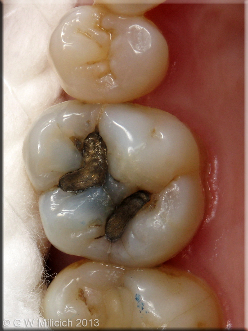
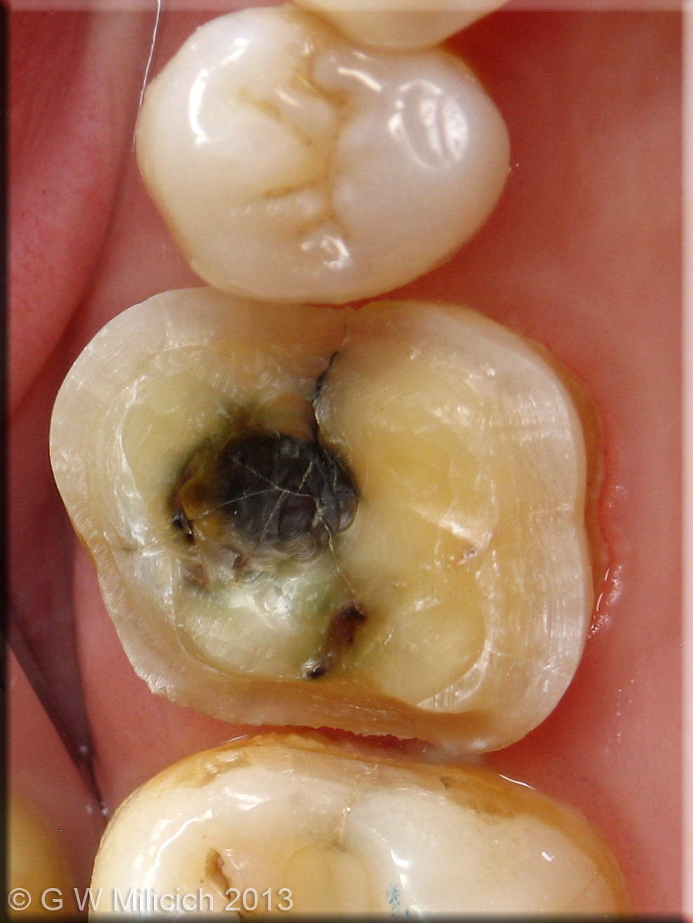
Certain dental procedures, such as tooth extractions, can provoke a condition known as osteonecrosis – or bone death – in patients taking biophosphonates, the most common treatment for osteoporosis. The drugs are also used as part of treatment for some cancers.

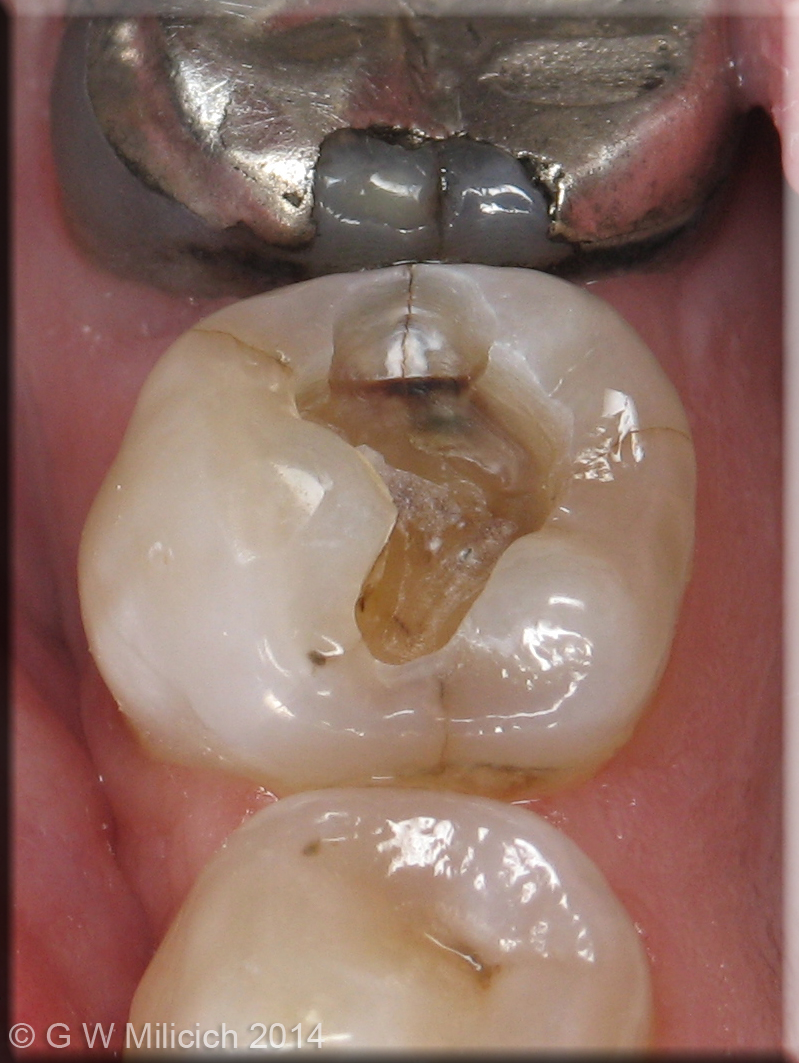
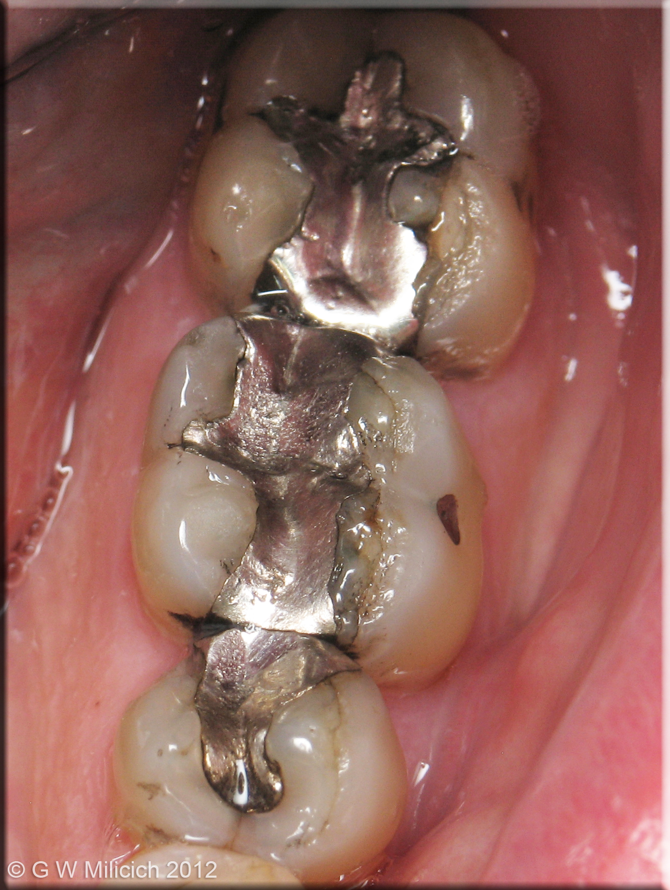
Osteonecrosis of the jaw is rare but is very difficult to treat and causes significant pain. Sometimes surgery is used to remove the dead bone, but this can be unsuccessful as there is a reduced blood supply to the affected area. The result can be disfiguring and often life-changing for patients.

A study into the impact of the disease on patients, carried out by a team from the University of Sunderland, found that much more communication was needed between GPs prescribing the biophosphonates, pharmacists supplying the medicines and dentists managing patient’s oral health.

Lead researcher Andrew Sturrock, Principal Lecturer and Programme Leader for the Master of Pharmacy programme at the University of Sunderland, explained: Although this condition is very rare, patients prescribed these medicines should have any dental treatment done before they start taking the drug. They also need to ensure they maintain good oral hygiene and good oral health whilst taking the medicine.

“Our findings, however, established that this wasn’t happening. GPs and pharmacists weren’t telling people about the risks for all sorts of reasons; because the disease is so rare, it wasn’t a priority or it’s been forgotten about. In some cases, the patient didn’t disclose they were taking the medicine when needing to have dental treatment.

Dentists are definitely out of the loop and they need to be more involved in the care of this patient group at a much earlier stage.”

The team interviewed 23 patients nationwide, six of whom had developed osteonecrosis as a result of taking biophosphonates. The remaining 17 participants all had been diagnosed with osteoporosis, of which thirteen had been prescribed biophosphonates.

The study, funded by Pharmacy Research UK and the UK Clinical Pharmacy Association, found that patients placed trust in their health professionals and expected that the risks and benefits of any medication should be communicated when the medicines were first prescribed. However, in most cases this didn’t happen, making it harder for patients to make informed decisions.

Many said their dentist did ask about drugs they were taken, but didn’t explain what the implications might be. All participants in the study said they would be happy for their information to be shared between health professionals, including communication between doctors and dentists when biophosponates were prescribed.

Co-author of the paper, Philip Preshaw, Professor of Dentistry at the University of Singapore, said: “Medication-related osteonecrosis of the jaw is thankfully quite rare, but when it does occur it has a profound impact on patients’ quality of life and can cause significant levels of pain.

“Prevention is always better than cure, and better communication between doctors, pharmacists and dentists would ensure that the dental team know which patients are going to start taking the implicated medications, so that they can assess and optimize their oral health before they start taking them, to reduce the risk of osteonecrosis developing later.”

Co-author Professor Scott Wilkes, Head of the School of Medicine, Professor of General Practice and Primary Care, University of Sunderland and part-time GP in North Tyneside, added: “GPs see a lot of patients who take bisphosphonate medications to protect against osteoporosis and Pharmacists dispense a lot of these medications every day.

“There are two opportunities missed for relatively simple interventions. The first is brief advice from the GP or Pharmacist to attend a dental check-up and the second is fully informed consent about the risk albeit uncommon. Pharmacy may have an opportunity to embed such a service into a New Medicines Service. Although rare, it is a devastating disease and any reduction in the number of cases is welcome.”

The research has now been published in the BMJ Open: https://bmjopen.bmj.com/content/9/3/e024376

The Six Fracture Modes of Teeth – Graeme Milicich – The British Dental Conference and Dentistry Show 2019

The Six Fracture Modes of Teeth

G W Milicich BDS

Teeth fracture for several reasons. It is very uncommon for a sound tooth to fail, but once a tooth has had a restoration placed, fractures begin to develop over a period of time. When we look very closely at how Mother Nature designed our teeth, several biomechanical strategies have been employed to ensure they can function for many decades. However, our modern diet and the associated decay epidemic, along with the required invasive management of the resultant decay disturbs the complex stress distribution system within a tooth. Effectively, a tooth behaves like a compression dome, similar to a cathedral dome. The enamel Bio-dome is designed to keep the underlying dentine in compression. When the enamel cap is disturbed to restore a tooth, important structures are damaged and this exposes the underlying dentine to tensile stresses it was not designed to cope with.  

There are several structures within enamel that work in strain harmony to ensure stability of the tooth. These include the sub-occlusal oblique transverse ridge, Rainey’s Web, the peripheral rim of enamel, occlusal enamel in general, and the Bio-rim (cervical half of the tooth). From a dentistry perspective, we should ideally be retaining these structures when restoring teeth. To achieve this, our primary gaol is to ensure retention and stability of the occlusal surface of the tooth. Minimally invasive dentistry’s primary aims are to manage a pathogenic biofilm and train it back to health. However, this is not always achievable, so protecting the fissure complex from damage is very important. This relies on being able to diagnose a pathogenic biofilm, or the early demineralization within a fissure complex, utilising all the modern technology we now have available, and predictably seal the fissures with autocure GIC. However, once the occlusal integrity has been disrupted, concentrated stresses within the underlying dentine lead to crack propagation and eventual failure of tooth structure. We must recognise enamel is not homogeneous, it is a very complex anisotropic structure, with amazingly subtle variations in ultrastructure and prism orientation, depending on the loads that have to be dissipated. 

There are six basic fracture modes within teeth.

The first two are associated with fractures in the peripheral rim of enamel.

Figure 1

Figure 2

  1. The first is described as “occlusal effect decay”. Once the occlusal enamel is cut, compression on a working cusp can cause distortion in the peripheral rim of enamel. This leads to an unstable vertical fracture where decay first become established at the EDJ and then work into the dentine and outwards to the surface of the enamel, following the internal walls of the vertical fracture. This fracture mode does not have an underlying dentine fracture.

Figure 3

  1. The second is a delamination of occlusal enamel and peripheral rim enamel beside an amalgam. Once again, the loss of the occlusal enamel allows the peripheral rim to flex under compressive forces, primarily on the working cusps. Investigating the complexities of the enamel cap, there is a distinct defining interface between occlusal and peripheral rim enamel that will delaminate if the peripheral rim enamel is flexing due to lack of support from the occlusal enamel. The adjacent amalgam provides no support to the surrounding enamel.

Figure 4

  1. Oblique cusp fractures. This is the most common fracture presentation that we see on a daily basis in our practices. Placing an amalgam into the occlusal of a tooth, that may or may not include either or both of the interproximal surfaces, exposes the underlying cuspal dentine to tensile forces. Over a period of years, a crack begins to propagate, eventually leading to the cusp fracturing off.

Figure 5 

Figure 6

Even though there is “only” an occlusal amalgam, this tooth has developed a mesio-distal fracture, as well as Poisson Effect fractures in the dentine underlying the amalgam.

  1. Mesio-distal fractures. This fracture mode is the nightmare of dentistry. Often diagnosis is difficult and symptoms are vague. This fracture mode is driven by vertical loads driving adjacent cusps apart, placing the underlying dentine into tension, but rather than the fracture generating obliquely, it travels vertically towards the pulp. Correct diagnosis and intervention can save most of these teeth.

(This tooth had all 4 cusps fail due to oblique fractures, but it also had multiple Poisson effect fractures)

  1. Poisson Effect Fracturing. A recently identified fracture mode that occurs under old amalgams. Once again, when the occlusal enamel is disrupted, the underlying dentine is exposed to tensile forces. Dentine is compressible and as a consequence is exposed to Poisson distortion. This can best be visualised as barrel distortion of the tooth. The overlying amalgam acts as a force concentrator, compressing the underlying dentine that then goes into radial tension. This leads to micro-fracturing of the dentine, creating random, often disconnected spiderweb-like fractures in the floor of the cavity. These can be present even in teeth with oblique or vertical fractures.
  2. Traumatic fractures. These are generally associated with either an external blow or an unexpected foreign object in food.

It is important that the fracture modality is accurately identified because management is quite varied. The primary goal is to identify where the forces that are causing the damage being applied to the tooth. If a crack is not dealt with correctly, the tooth can continue to exhibit symptoms and often a secondary diagnosis of an irreversible pulpitis is made, leading to unnecessary endodontics. If the fracture is correctly diagnosed and treated, the tooth generally settles.

As an overview, the goal is to re-create a stable compression dome to prevent the underlying dentine from being exposed to tensile forces. With more damaged teeth, this is best achieved with an adhesive ceramic onlay. However, depending on the fracture type, many teeth can be predictably stabilised using direct bonded composite in conjunction with Ribbond fibre reinforcing. Success relies on being able to predictably bond to tooth structure in the long term. Some in our profession do not believe this is possible, but clinical success for over 30 years using the Biomimetic techniques described in my lecture in the BDA Theatre will prove it is not only possible, but totally predictable. Once we come to an understanding of how a tooth functions at a biomechanical level, it becomes easier to diagnose the various failure modes of teeth and then predictably restore them. Equally, the less you do to a tooth in the beginning, the less you will have to do to it in the future.

When a minimally invasive philosophy is adopted, and biomimetic restorations become an option, dentists notice a significant change in their practices. The incidence of post treatment endodontics is reported by Biomimetic dentists to reduce by 80-90%, because they are diagnosing fractures accurately and treating them appropriately. 

Dr Graeme Milicich will be discussing the topic in further detail within the BDA Theatre at the British Dental Conference and Dentistry Show 2019. Register for your free delegate pass to make sure you don’t miss out.


The British Dental Conference and Dentistry Show 2019 will be held on Friday 17thand Saturday 18thMay at the Birmingham NEC, co-located with DTS.


Visit www.thedentistryshow.co.uk, call 020 7348 5270

or email dentistry@closerstillmedia.com


Author biography:

Graeme Milicich

Graeme is a graduate of the University of Otago (Dunedin, NZ) and maintained a private general practice in Hamilton since 1977 (NZ) until 2017 when he retired from active practice. Throughout his career he developed a keen interest in the area of minimal intervention dentistry (MID), which was considered in decades past to be revolutionary. This area of interest quickly drew his attention to caries risk management, biomechanics and biomimetics as it applied to restorative dentistry – lasers and CAD/ CAM and their applications in minimally invasive dentistry have been at the forefront of Graeme’s expansion of MID concepts.  

He has several peer-reviewed published articles and has presented at a national and international level in the field of MID, been recognised by several international bodies dedicated to furthering MID and laser dentistry and has been a clinical educator in CAD/CAM dentistry. Graeme has developed many training resources in the field of MID and undertaken clinical studies into the application of lasers for restorative dentistry.


Time to switch?

As one of Britain’s leading suppliers of dental consumables and equipment, Denka stocks over 1,000 products for your general and specialist needs.

If you are looking for protective wear, Denka offers a comprehensive range of solutions – from aprons and gowns to eyewear, facemasks and patient bibs. All the options are not only high quality to optimise patient and practitioner safety, but with different pack sizes for many of the products, they represent great value.

With nearly two decades in the dental sector and an unwavering commitment to a customer-focused approach, is it time that your practice switched to Denka? Request the latest catalogue today!

For more information, visit www.denkauk.com, call on 0800 707 6212 or email support@denkauk.com

Learn something new

Join hundreds of delegates at the British Dental Conference and Dentistry Show 2019 for a fantastic line-up of engaging lectures and hands-on workshops. The BACD’s Aesthetic and Digital Dentist Theatre will return with the ideal stage to provide information on the latest trends within the cosmetic dental profession.

Members of the Academy will also be on-hand to discuss the benefits of BACD membership – including networking opportunities, unlimited access to exciting educational events, and the chance to join the prestigious Accreditation Programme.

Be sure to come and meet the BACD at the British Dental Conference and Dentistry Show in May – you won’t want to miss out!


For further enquiries about the British Academy of Cosmetic Dentistry, visit www.bacd.com

Laying the foundations – Kate Scheer W&H UK

The latest dental implant systems offer various benefits, perhaps the most important of which is achieving a stable, natural-looking replacement for missing teeth. Implants have consequently become a highly accepted treatment modality for partial or complete edentulism, with many patients now choosing implants over traditional alternatives such as dentures or bridges. However, the placement and restoration of an implant requires meticulous assessment and evaluation to ensure its long-term success. In particular, the condition of a patient’s jaw bone is key to achieving primary stability, which is vital to successful osseointegration.[i]

One of the challenges that practitioners face when planning for dental implant treatment is the extent of bone resorption following tooth loss. As practitioners know, natural teeth are supported and attached to the jaw bone via the periodontal ligament (PDL), which acts as a shock absorber to distribute occlusal forces and stimulate the bone. In the case of tooth loss or extraction, the bone that once surrounded the missing tooth gradually deteriorates due to a lack of stimulation. In fact, the rate of bone resorption is highest within the first six months following extraction, and proceeds at an average of 0.5% to 1.0% each year throughout an individual’s lifetime.[ii]

Although the degree of bone resorption varies from patient to patient, it will always occur to some extent unless specific care is taken to avoid it. This remains an issue even for a denture wearer, whose appliance is not in direct contact with bone and therefore doesn’t stimulate it effectively. Over time, it is the effect of mastication that causes gradual deterioration of the bone structure supporting the denture, which could alter the fit of the appliance and detrimentally affect its stability.[iii]In contrast, a dental implant is designed to mimic the function of natural tooth roots and remains stable following osseointegration with the jaw bone. Essentially, the longer a tooth is missing, the less bone is available to safely place an implant into.

In this case, bone grafting procedures may be required to ensure that the patient has adequate bone quality and quantity, which is crucial to determining the clinical success of implant treatment.iBone grafts can be carried out at the same time or prior to placing an implant. It is evidently more convenient to place an implant and perform a bone graft simultaneously – thus reducing the time required for surgery – but the clinical situation might not always allow for this. There is a variety of different bone grafting procedures that practitioners can perform and selection will depend on each case. Ultimately, the time, effort, and additional expense of successfully performing a bone graft can greatly improve the aesthetic and functional outcome of an implant – particularly if a patient is suffering from extensive bone resorption.

Once an implant is placed, everyday masticatory forces will begin to stimulate the surrounding bone, which responds by becoming stronger and denser.[iv]Yet, there is a range of external factors that can influence the future preservation of the bone. Slight bone loss over several years is considered normal and should not affect the patient’s implant directly. However, implant treatment is deemed a failure if there is bone loss of more than 1.5mm following the first year of placement, and more than 0.2mm every year after.[v]Patients who suffer from diabetes, osteoporosis, or a poor immune system are at greater risk of bone loss as they already have weak bones.[vi]

Similarly, patients with a history of periodontal disease – typically as a result of poor oral hygiene – are more likely to contract peri-implantitis, which is a common cause of dental implant failure.[vii]This inflammatory disease can damage the gingiva if left untreated, resulting in deterioration of the bone structure supporting the implant. Smokers, in particular, are in danger of developing peri-implantitis because nicotine is a vasoconstrictor that reduces blood flow, causing tissue ischemia and impaired healing of injured tissue.[viii]This can negatively impact the condition of the bone, especially in the initial stages of post-surgery healing and in the case of regeneration if a bone graft has been performed.

Essentially, without a solid foundation in which to place an implant, clinicians are unable to achieve primary stability. This emphasises the need for advanced diagnostic tools that can help guide practitioners in optimising implant treatment for both simple and complex cases. As one of the industry’s leading manufacturers, W&H offers an innovative way to do this through the Osstell range of products, including the new Osstell Beacon. This intuitive handheld device facilitates a non-invasive way of measuring primary implant stability, observing osseointegration based on secondary stability readings, and aiding practitioners in determining the best possible time for implant loading. For those seeking a complete system, the Implantmed surgical unit is available to purchase with the Osstell ISQ module already integrated. Consequently, clinicians are able to prevent implant failure, reduce healing time, and ensure the quality of treatment for highly effective, long-term results.


To find out more visit www.wh.com/en_uk, call 01727 874990 or email office.uk@wh.com


References

[i]Javed, F., Ahmed, H. B., Crespi, R., and Romanos, G. E. (2013) Role of primary stability for successful osseointegration of dental implants: Factors of influence and evaluation. Interventional Medicine & Applied Science. 5(4): 162–167. Link: http://doi.org/10.1556/IMAS.5.2013.4.3. [Last accessed: 19.09.18].

[ii]Pagni, G., Pellegrini, G., Giannobile, W. V. and Rasperini, G. (2012) Postextraction Alveolar Ridge Preservation: Biological Basis and Treatments. International Journal of Dentistry. 151030. Link: https://www.ncbi.nlm.nih.gov/pmc/articles/PMC3378971/#B8. [Last accessed: 19.09.18].

[iii]Knezović-Zlatarić, D., Čelebić, A. and Lazić, B. (2002) Resorptive Changes of Maxillary and Mandibular Bone Structures in Removable Denture Wearers. Acta Stomat Croat. 261-265. Link: https://hrcak.srce.hr/file/6144. [Last accessed: 19.09.18].

[iv]Association of Dental Implantology. (2018) Bone – the foundation for dental implants. Link: http://consideringdentalimplants.co.uk/considering-dental-implants/bone.html. [Last accessed: 19.09.18]. 

[v]Albrektsson, T., Zarb, G., Worthington, P. and Eriksson, A.R. (1986) The long-term efficacy of currently used dental implants: a review and proposed criteria of success. Link: https://www.ncbi.nlm.nih.gov/pubmed/3527955.[Last accessed: 19.09.18]. 

[vi]Chen, H., Liu, N., Xu, X., Qu, X. and Lu, E. (2013) Smoking, Radiotherapy, Diabetes and Osteoporosis as Risk Factors for Dental Implant Failure: A Meta-Analysis. Link: http://journals.plos.org/plosone/article?id=10.1371/journal.pone.0071955. [Last accessed: 19.09.18].

[vii]Prathapachandran, J. and Suresh, N. (2012) Management of peri-implantitis. Dental Research Journal. 9(5): 516-521. Link: https://www.ncbi.nlm.nih.gov/pmc/articles/PMC3612185/. [Last accessed: 19.09.18].

[viii]Kasat, V. and Ladda, R. (2012) Smoking and dental implants. Journal of International Society of Preventive & Community Dentistry. 2(2): 38-41. Link: https://www.ncbi.nlm.nih.gov/pmc/articles/PMC3894084/. [Last accessed: 19.09.18].

Even the Tooth Fairy is feeling the pinch – payments are down by 10% to an average of £1.90 per gnasher, according to a new survey

Payments from the Tooth Fairy are down by 10% to an average of £1.90 per gnasher, according to a new survey.

A small minority of youngsters (2%) get as much as £20 for each tooth that falls out – amounting to £400 for a full set of all 20 baby teeth.

But average payments across the UK are down.

London and the South-East has the highest figure with an average of £2.25 a tooth – down 10% from £2.50 four years ago.

The Tooth Fairy is most careful in Newcastle where kids get an average of 80p per lost tooth – a 20% slump on 2015’s figure.

The results came from a new survey of 2,000 parents by cosmetic dental brand Your Smile Direct.

The survey found that average payments nationally had dropped by 10% from £2.10 to £1.90 since 2015.

More than a quarter (28%) of children get a £1 coin for each lost tooth, 24% get a £2 coin, and 14% get less than £1 – most typically 50p.

A further 11% get £5, 10% get £10, 3% get between £10 and £20 and 2% get more than £20.

Only 8% of kids never receive a visit from the Tooth Fairy.

Nine out of ten parents (91%) said their children under five believed in the Tooth Fairy – the same figure as for Santa Claus.

More than a third of parents (34%) said their children spent their Tooth Fairy money on sweets.

A further 33% spent it on toys, with saving (21%), books (7%) and clothes (5%) the other most popular answers.

Dr Mark-Anthony Shepherd, lead dentist for Your Smile Direct, said: “Even the Tooth Fairy is feeling the pinch!

“But it is great news that the Tooth Fairy is still visiting 92% of homes with children.

“It is a shame that a third of children are spending their Tooth Fairy money on sweets – that is the quickest way to ensure a further visit from the Tooth Fairy.”

Most children have a full set of 20 milk or baby teeth by the age of three and start losing them by the age of five or six. They tend to fall out in the same order they came, with the front centre lower teeth going first.

It takes six or more years to grow a full set of 28 adult teeth – 32 if you include wisdom teeth which arrive right at the back of the mouth around the age of 20.

Your Smile Direct offers dentist prescribed invisible aligner treatment which patients use at home – removing the need for repeated, costly dental visits.

It costs £1,599 – around a quarter of the price of teeth straightening with conventional dentists.

Average payments from the Tooth Fairy across the UK

1 London and the South-East  – £2.25

2 Bristol  – £1.70

3 Aberdeen £1.68

4 Cardiff  – £1.64

5 Birmingham £1.58

6 Leeds – £1.46

7 Manchester – £1.40

8 Belfast £1.34

9 Edinburgh £1.28

10 Coventry £1.26

11 Leicester £1.22

12 Nottingham £1.18

13 Plymouth £1.15

14 Sheffield £1.12

15 Liverpool £1.05

16 Sunderland £1

17 Glasgow  95p

18 Bradford 91p

19 Wakefield 85p

20 Newcastle 80p

What do children buy with their Tooth Fairy money?

1 Sweets 34%

2 Toys 33%

3 They save it 21%

4 Books 7%

5 Clothes 5%

Most popular payments from the Tooth Fairy

1 £1 28%

2 £2 24%

3 Less than £1 – most typically 50p 14%

4 £5 11%

5 £10 10%

6 Nothing 8%

7 Between £10-£20 3%

8 More than £20 2%

A lab partner you can trust

In order to meet patients’ growing expectations and ensure their satisfaction with treatment, it’s essential to use high quality materials.

It is equally as important that your practice utilises quality solutions and this is where Sparkle Dental Labs could help.

Dedicated to providing an array of high quality services, the team use only clinically proven and reputable products. Combined with cutting-edge technologies such as 3D printers and digital scanners, this enables Sparkle Dental Labs to produce accurate and consistent quality restorations for a range of indications.

For confidence that you can deliver optimal treatment results for your patients, work with a lab partner you can trust.


For any additional information please call 0800 138 6255 or email customerservice@sparkledentallabs.com or visit:

www.sparkledentallabs.com

Empowering women

“The best aspect of the BACD’s Ladies Who Do Dentistry event is that it empowers women to follow their own career paths, form supportive networks, and help them realise that they can speak to other women who have been successful within the dental profession,” says Dr Ewa Rozwadowska.

“I think it’s fantastic that the BACD is embracing this initiative. I hope the Academy has many more delegates attending the upcoming Ladies Who Do Dentistry event on Saturday the 23rd of March 2019. One of the speakers at this event will explore the creation of wealth and financial planning for women – especially at the start of their careers.”

For further enquiries about the British Academy of Cosmetic Dentistry, visit www.bacd.com

Combatting biofilms – Arifa Sultana

The oral cavity has many surfaces, each coated with hundreds of species of bacteria. Some bacteria are free-floating but for the most part, they multiply and flourish within a complex living community or biofilm. Here they are held together and nourished in a self-supporting network, which attaches and adheres to surfaces and is protected by an outer matrix or extracellular slime layer that is extremely resistant to antibiotics, antimicrobials and human defence mechanisms.[1],[2]

Dental plaque is recognised as a biofilm that adheres to tooth surfaces, restorations, dental implants and prostheses. It is present in the oral environment of healthy individuals but if left to accumulate, the teeth and gingival tissues may be subjected to high concentrations of bacterial metabolites, which can result in dental disease and decay. It is notoriously difficult to completely destroy biofilms and as dental professionals are aware, continuous and regular disruption is the only effective method for controlling biofilm growth, maintaining oral health and preventing oral disease progression.

Biofilms also form in the sinuses and adenoids, at the back of the mouth cavity and behind it, for example in the crypts at the base of the tongue, around the pharynx and on the palatine tonsils. Interestingly, it has recently been suggested that as well as offering first line defence against viral, bacterial, and food antigens, the palatine tonsils may also influence the growth and control of bacteria.[3],[4]Certainly, these soft tissues have natural folds and pockets where debris such as dead cells, food particles, mucus and large concentrations of bacteria can become trapped, and if left undisturbed, can form calcified concretionscalledtonsilloliths or tonsil stones.

In general, tonsil stones are considered to be fairly harmless. Often they are only detected incidentally on X-rays or CT scans and do not cause any noticeable symptoms. Some tonsilloliths, however, can appear as white or cream coloured lumps on the soft tissues at the back of the throat, which may cause irritation or the feeling that there is a constant obstruction in the throat. Depending on their size and location tonsil stones can also cause difficulty or pain when swallowing and some patients may experience referred pain in the ear. Often patients report a metallic taste in the mouth and symptoms vary from individual to individual. Nevertheless, tonsilloliths are living biofilms comprising of a significantly high number and density of bacteria and most patients with tonsil stones experience extremely unpleasant smelling breath.[5]

Tonsil stones comprise of large amounts of anaerobic bacteria including Eubacterium, Fusobacterium, Megasphaera, Porphyromonas, Prevotella, Selenomonasand Tannerella, which according to research, all appearto be associated with the production of volatile sulphur compounds.[6]To explain,

volatile sulphur compounds, or VSCs, are foul smelling gases formed as by-products during the interaction and microbial putrefaction of food debris, cells, saliva and blood, which are expelled in the breath.[7]Primarily, VSCs are composed of hydrogen sulphide, which has the odour of rotting eggs, methyl mercaptan with a barnyard smell and dimethyl sulphide, which smells of rotting cabbage or garlic.

Research has revealed that tonsilloliths are 10 times more likely to produce elevated levels of VSCs in the breath.5 Therefore, to effectively prevent the build up of bacterial biofilm and the production of VSCs, patients need to implement a regular and thorough oral hygiene routine with brushing and flossing to physically disrupt bacterial growth, and a specifically prepared mouthwash containing Chlorhexidine (CHX).CHX is bacteriostatic (inhibits bacterial growth) and bactericidal (kills bacteria). It works rapidly and binds to the oral tissues, allowing its antimicrobial effects to be sustained for several hours. In low concentrations CHX is able to damage bacterial cell walls, attack the inner membranes causing component leakage and, subsequently, cause cell death.[8]It can inhibit the adherence of microorganisms to a surface and prevent the growth and development of biofilms.

CB12 mouthwash was developed by dentists and has a patented formula containing CHX and zinc acetate, which specifically targets and neutralise foul smelling oral gases. This mouthwash successfully converts the offensive sulphur content of VSCs into odourless, insoluble sulphides. The active ingredients in CB12 are also able to chemically bind to the tissues of the oral cavity where it continues to neutralise gases and prevent oral malodour for up to 12 hours.[9]In addition, CB12 mouthwash contains fluoride to help strengthen the teeth and prevent dental decay.

Destroying problematic biofilms within the oral cavity can be challenging. However, these microbial communities can be inhibited considerably by physical cleaning and rinsing away bacteria and debris as often as possible. The emphasis is on prevention, with a good oral hygiene routine and the use of effective, clinically proven oral health products.


For more information about CB12 and how it could benefit your patients, please visit www.cb12.co.uk


References

[1]Nield-Gehrig J.S, RDH, MA Dental Plaque Biofilms. http://bjjcaveman.com/wp-content/uploads/2015/07/Denta-Plaque-Biofilms.pdf[Accessed 6th November 2018]

[2]Jamal M. et al. Bacterial biofilm and associated infections. Journal of Chinese Medical Assoication. 81 (2018) 7-11. https://reader.elsevier.com/reader/sd/pii/S1726490117302587?token=DCB218C23962F03283C9956C4FD4149258477E5879BFDF22D0DEDDB55EF6B62EEF2A4A77CD78874468CC77CF369745F1[Accessed 6thNovember 2018]

[3]Bernstein J.M. Mucosal Immunology of the Upper Respiratory Tract.

Respiration 1992; 59:3–13. https://www.karger.com/Article/Pdf/196123[Accessed 6th November 2018]

[4]Weise J.B. et al. A newly discovered function of palatine tonsils in immune defence: the expression of defensins. The Polish Ontolaryngology. 2002 56(4) 409-413.http://mbbsdost.com/Weise-JB-et-al-2002-/et-al/14582658[Accessed 6th November 2018]

[5]Ferguson M. et al. Halitosis and the Tonsils: A Review of Management . Otolaryngol Head Neck Surg 2014, 151(2): 0194599814544881. https://pdfs.semanticscholar.org/d6de/09ea629a648a356e35453b72089af6995d21.pdf[Accessed 6th November 2018]

[6]Tsuneishi M. et al. Composition of the bacterial flora in tonsilloliths. Microbes Infect. 2006 Aug;8(9-10): 2384-9.https://www.ncbi.nlm.nih.gov/pubmed/16859950. {Accessed 6th November 2018]

[7]Uğur Aylıkcı B. et al. Halitosis: From diagnosis to management. J Nat Sci Biol Med. 2013 Jan-Jun; 4(1): 14–23. https://www.ncbi.nlm.nih.gov/pmc/articles/PMC3633265/[Accessed 6th November 2018]

[8]Chlorhexidine Facts. About Chlorhexidine: Mechanism of Action.  http://chlorhexidinefacts.com/mechanism-of-action.html[Accessed 6th November 2018]

[9]Thrane PS et al. Zn and CHX mouthwash effective against VSCs responsible for halitosis for up to 12 hours. Journal of the British Society of Dental Hygiene and Therapy, Dental Health Vol 48 2009 No 3 of 6. http://www.cb12.fr/fileadmin/user_upload/cb12_fr_new/pdf/studie_090929_2.pdf[Accessed 6th November 2018]


A new dimension in dental fabrication – Stephen Norman – Private Implant prosthetics Manager at Sparkle Laboratory

While 3D printing has been hailed as a disruptive technology, with applications ranging from the artistic to robotics, it actually has its root in early 1980s additive manufacturing. However, it is only recently that the technology has become mainstream, thanks to advances in accessibility and technique.

Being able to quickly and relatively cheaply go from concept to prototype to a finished piece is truly an exciting development. Doctors have been able to fabricate bespoke prostheses and implants as needed, such as creating a cranial implant from PEEK.[i]The benefits aren’t even limited to the terrestrial! Launching any payload into space is infamously costly due to the quantity of rocket fuel required. Previously, if for instance an astronaut on the International Space Station lost a tool while on a spacewalk, they would have to wait for a replacement to be sent, which could take months. That tool would take up precious space on the next resupply run – which can cost $43,180 per pound![ii]Armed with a 3D printer, astronauts can now potentially replace some parts and equipment themselves, making them far less dependent on resupply than in the past.[iii]

Key advantages

Speed and flexibility. From prototype to finished product, 3D printing can streamline fabrications. It is not unknown for those with access to 3D printers to quickly throw together a simple design that fulfils a specific need on the spur of the moment. Rapid prototyping enables 3D printers to produce everything from orthodontic appliances to maxillofacial prostheses.[iv]

Minimal waste. Traditionally, subtractive manufacturing techniques have been employed, which are inherently more wasteful of component materials by their very nature.[v]Quite simply, when building up the operator can use just the material required by the design, baring a few small excesses that may need to be sanded or washed off. This is markedly in contrast to subtractive methods where excess material must be removed from the construct.

Dental applications

As in so many areas, dentistry is now benefiting from 3D printing too. For instance, rapid prototyping methods can produce accurate anatomical models from scans of an individual. These can greatly aid oral surgeons, as rather than only having visual information prior to an operation, that can now be augmented with tactile replicas of the patient’s physiology to help plan the procedure. Another usage for such facsimiles is as a learning aid for dental students, or to illustrate a point to a patient.[vi]

3D printing is contingent on Computer Aided Design (CAD). CAD allows for complex three-dimensional objects to be modelled digitally. This can be done from scratch, however, many dental practices already have access to volumetric data, captured by CBCT machines and the like. This data can be fed directly into certain programs, enabling the swift creation of a highly accurate digital model of the region scanned. Once successfully imported and adjusted, this data can be used for diagnostic purposes. However, when combined with 3D printing this allows for the creation of bespoke implants and surgical guides.[vii]But just because a computer is involved doesn’t mean everything is automatically done for you. While much of the process is automated, a skilled CAD operator is still required.

The advances and increasing availability of these two synergistic technologies benefit dentists, who can not only gain valuable diagnostic information from CBCT scans but also use the data to have surgical guides and prostheses produced to exacting specifications.

Selective laser sintering

3D printing isn’t just restricted to plastics and epoxies, alloys can be used too. Porous titanium implants can be 3D printed by using a high-powered laser to fuse the metal particles on a powder bed, layer by layer.[viii]However, to make full use of polymers and metals requires equipment beyond the type of additive 3D printers found in the homes of enthusiasts. For these materials technologies like selective laser sintering (SLS) are required, which is currently prohibitively expensive and dangerous outside of specialist hands, due to the risk of dust inhalation or even an explosion.[ix]Directly printing in metal can require substantial post-processing before the piece(s) are suitable for clinical use.[x]

It should be noted that ceramics, which see widespread use in dentistry, cannot be printed using any existing methods, so there are still some limitations to what 3D printers can produce.

A skilled laboratory

As exciting as the prospects of 3D printing are, the cost and expertise required to make proper use of the technology can be prohibitive – especially if you require certain materials like metals to be used. 

Sparkle Dental Labs is a full service in-house laboratory offering reasonable prices and an efficient turn around. It is well equipped to offer 3D printing, laser sintering and milling – being the very first dental lab in the UK to use both the Renishaw AM250 Laser Sintering Machine and DryLyte Chrome Polishing System. Whatever your practice needs, our technicians are ready, willing and able.

3D printing is an exciting technological addition to dentistry, bringing a flexible and speedy means of production to the field. From prosthetics to treatment planning there are already many applications for 3D printing, it is sure to be part of dentistry’s future – one you can take advantage of today.


For any additional information please call 0800 138 6255 or email customerservice@sparkledentallabs.comor visit:

www.sparkledentallabs.com


References

[i]Honigmann P., Sharma N., Okolo B., Popp U., Msallem B, Thieringer F. Patient-specific surgical implants made of 3d printed PEEK: material, technology, and scope of surgical application. BioMed Research International.  2018; 2018: 4520636. https://www.ncbi.nlm.nih.gov/pmc/articles/PMC5884234/Accessed November 30, 2018.

[ii]Kramer S. It still costs a staggering amount of money to launch stuff into space. Business Insider UK. July 20, 2016. http://uk.businessinsider.com/spacex-rocket-cargo-price-by-weight-2016-6Accessed November 30, 2018.

[iii]Prater T., Bean Q., Beshears R., Rolin T., Werkheiser N., Ordonez E., Ryan R., Ledbetter III F.  Summary report on phase I results from the 3D Printing in Zero G technology demonstration mission, volume I. NASA Technical Publication. 2016.https://ntrs.nasa.gov/archive/nasa/casi.ntrs.nasa.gov/20160008972.pdfAccessed November 30, 2018

[iv]Nayar S., Bhuminathan S., Bhat W. Rapid prototyping and stereolithography in dentistry.  Journal of Pharmacy & Bioallied Sciences.2015; 7(Suppl. 1): 216-219. https://www.ncbi.nlm.nih.gov/pmc/articles/PMC4439675/Accessed November 29, 2018.

[v]Prasad S., Kader N., Sujatha G., Raj T., Patil S. 3D printing in dentistry. Journal of 3D Printing in Medicine.2018; 2(3): 89-91. https://www.futuremedicine.com/doi/full/10.2217/3dp-2018-0012Accessed November 29, 2018.

[vi]Zaharia C., Gabor A., Gavrilovici A., Stan A., Idorasi L., Sinescu C., NegruțiuM. Digital dentistry – 3d printing applications. Journal of Interdisciplinary Medicine. 2017; 2(1): 50-53. https://content.sciendo.com/abstract/journals/jim/2/1/article-p50.xmlAccessed November 30, 2018.

[vii]Dawood A., Marti B., Sauret-Jackson V. 3D printing in dentistry. British Dental Journal. 2015; 219(1): 521-529. https://www.researchgate.net/publication/286612886_3D_printing_in_dentistryAccessed November 29, 2018.

[viii]Prasad S., Kader N., Sujatha G., Raj T., Patil S. 3D printing in dentistry. Journal of 3D Printing in Medicine.2018; 2(3): 89-91. https://www.futuremedicine.com/doi/full/10.2217/3dp-2018-0012Accessed November 29, 2018.

[ix]Prasad S., Kader N., Sujatha G., Raj T., Patil S. 3D printing in dentistry. Journal of 3D Printing in Medicine.2018; 2(3): 89-91. https://www.futuremedicine.com/doi/full/10.2217/3dp-2018-0012Accessed November 29, 2018.

[x]Dawood A., Marti B., Sauret-Jackson V. 3D printing in dentistry. British Dental Journal. 2015; 219(1): 521-529. https://www.researchgate.net/publication/286612886_3D_printing_in_dentistryAccessed November 29, 2018.