


-

‘Lifting my practice to the next level’
Struggling to decide upon a CBCT unit for his new practice, Dr Martin Sulo received a strong... -
From Helsinki to Nottingham
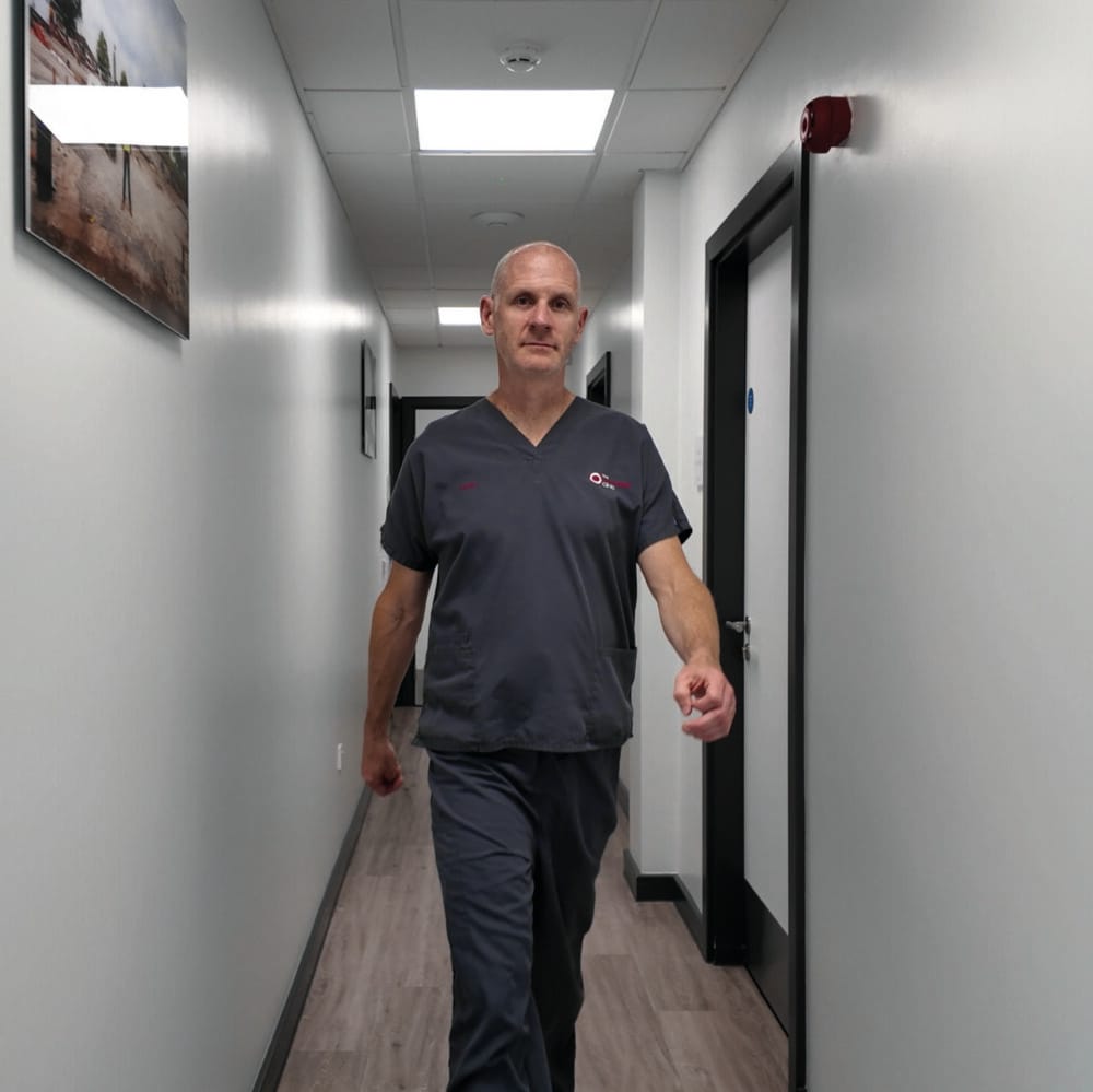
Colin Campbell, Director at The Campbell Clinic, recalls how Planmeca has supported him in getting...


