Dr Hamed Safaei presents a case in which he placed a dental implant following a fully guided, immediate placement protocol, achieving excellent results.
Introduction
When a tooth is deemed to have a hopeless prognosis, there is a myriad of treatment options that present themselves. Research has found that the use of a fully guided surgical technique results in highly accurate implant positioning in fresh extraction sites when compared to a partially guided approach.[i] It is important to reduce deviations when placing implants in tight spaces between teeth, in order to reduce the risk of future complications. In particular, the predictability provided by guided protocols in ensuring the correct angle is achieved might be considered the most critical parameter, offering long-term peri-implant tissue stability and implant survival.i
Patient presentation and assessment
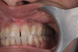
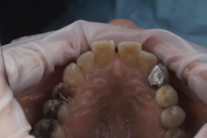
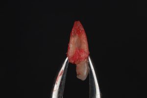
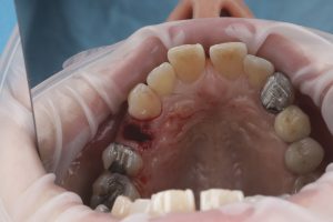
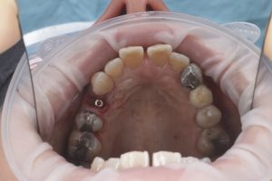
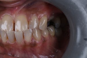
In this case, the patient was a 60-year-old lady. Her UL4 tooth had snapped in such a way that it could not be saved. Otherwise, she had a heavily restored dentition with a number of fillings, crowns, and bridges already in place, and no relevant medical background.
Initially, visual confirmation was obtained through a clinical exam, with CBCT used to confirm that there was no infection present, but that the tooth had snapped in a way that it could not be saved anymore and that there was enough bone volume above the apex of the tooth for an immediate implant placement. These tests helped to confirm the suspected diagnosis, that the tooth had a hopeless prognosis and it was a restorative failure.
Treatment planning
All treatment possibilities were discussed as part of planning for this case. This included extracting the problem tooth and leaving a gap, or restoring with a cantilever bridge attached to the already heavily restored UL5. Whilst each of these were technically viable options, they were not preferred, with the patient seeking a long-term solution. Each of these options would have allowed the bone to recede over time, leading to more complex issues down the line.
As such, the third option was to provide a single dental implant at the premolar site. It was a nice site for an implant, and the patient had already received two implants before, so was very comfortable with consenting to this option, and clearly understood what to expect.
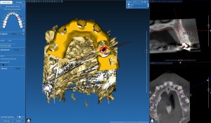
Initially, an intraoral scan was taken, and merged with the CBCT scan using RealGuide software. From this, the future position of the implant was planned and a surgical guide was designed. The surgical guide was then created by an external dental lab (Go2Guide).
Treatment provision
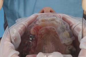
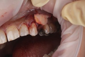
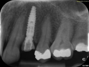
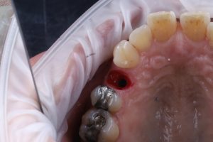
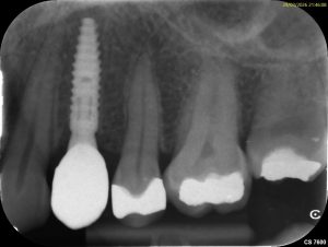
To begin with, the broken tooth was extracted, then a pre-planned drill sequence was followed through the surgical guide, and a 3.3mm x 13 CONELOG® Progressive-Line implant was placed using a fully guided protocol. I regularly utilise the CONELOG® Progressive-Line implant as I have seen good long-term results and felt it was ideal for use in this clinical situation.
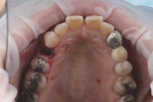
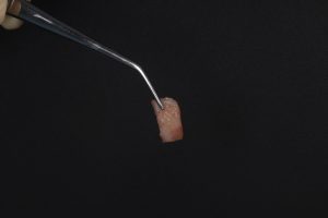
Following implant placement, MinerOss® Cortico-Cancellous Blend bone graft was placed on the buccal side to close the jump gap. I use MinerOss® Blend frequently, having initially been recommended it by Dr Rob Oretti many years ago, and get great results. Following this, connective tissue graft was harvested from the palate and placed on the buccal side also to ensure sufficient soft tissue volume.
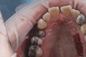
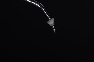
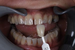
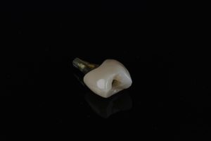
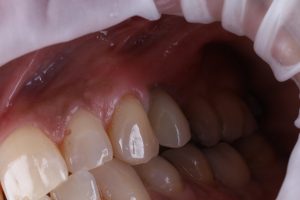
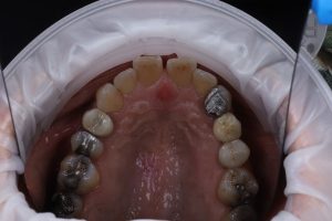
A custom healing abutment was made and provided on the same day, and left in place for three months. At this stage, good healing was observed and a permanent restoration was provided, created by a dental lab (Digital Prosthetics).
Case appraisal
Both myself and the patient were very happy with the treatment results, which have shown excellent aesthetic and functional outcomes, giving her a stable and long-lasting solution.
In order to maximise the chances of success in a case like this, there are a number of things that must be taken into consideration. Careful case selection is essential for immediate implant placement: Adequate apical bone beyond the extraction socket, absence of acute infection, and the ability to achieve primary stability should be confirmed before proceeding. Further to this, extraction technique is critical. Atraumatic extraction and preservation of the buccal plate are fundamental to maintaining hard and soft tissue architecture around immediate implants. Finally, the use of custom healing abutments can aid tissue shaping from day one. Immediate contouring of the peri-implant soft tissues may simplify the restorative phase and improve the final aesthetic outcome.

For product information or further education from BioHorizons and Camlog, please visit https://theimplanthub.com/
 Author Bio: Dr Hamed Safaei is an implant and cosmetic dentist, passionate about restoring patients’ smiles. Providing everything from single unit to full-arch solutions, his precision and artistry unite to deliver long-lasting, natural-looking results. Meticulous planning and patient-centred care are key, utilising digital dentistry and guided protocols to make treatment predictable.
[i] Nicchio, N., Gonçalves, V., Mendonça, G., e Pessoa, R.S., Frizzera, F. and Zandim-Barcelos, D.L., 2023. Accuracy of partially and fully guided surgical techniques for immediate implant placement: An in vitro assessment. The Journal of Prosthetic Dentistry, 129(2), pp.363-e1.