





 Author: Dr Farnam Pourreza-Jorshari BDS PGDip, British Endodontic Society Early Career Group Committee Member
Historically, pulp necrosis has been managed with root canal treatment (RCT), in which diseased pulp tissue is removed and replaced with an inert filling material, permanently sacrificing pulpal function in the process. Regenerative endodontic techniques (RETs), also termed revascularisation or revitalisation, seek instead to restore the vital pulp within the necrotic canal system. This is clinically meaningful: the dental pulp performs formative, defensive and sensory functions that conventional RCT cannot replace. Beyond root development and dentine deposition, the pulp-dentine complex provides immune defence, detects noxious stimuli and confers proprioception.[i] This article reviews the evolution, biological principles and clinical evidence underpinning RETs, while examining the biological barriers that continue to limit predictable pulp-dentine regeneration.
Nygaard-Østby[ii] first demonstrated that a blood clot could be induced within instrumented root canals following chemo-mechanical debridement and enlargement of the apical foramen, resulting in resolution of infection and apical narrowing. A subsequent histological study by Nygaard-Østby and Hjortdal[iii] found the new intracanal tissue resembled calcified connective tissue rather than true pulp, but nevertheless established that the canal space could be biologically repopulated. Case reports by Iwaya et al.[iv] and Banchs and Trope[v] subsequently built upon this concept, culminating in a European Society of Endodontology (ESE) position statement recognising RETs as a viable treatment modality.[vi]
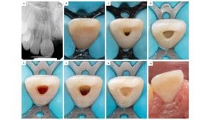
Although single-visit protocols have been described[vii],[viii], the ESE recommends a two-visit approach involving minimal instrumentation, irrigation with low-concentration sodium hypochlorite and EDTA, intracanal medication, induction of apical bleeding, placement of a calcium silicate cement and definitive adhesive restoration[ix] (Figure 1).
Successful regeneration depends on three core components: stem cells, signalling molecules and a scaffold. In RETs, the blood clot serves as a scaffold, while dentine stores growth factors (IGF-1, TGF-β1, BMP-2, BMP-4, BMP-6 and GH), released following demineralisation by EDTA, calcium hydroxide or calcium silicate cements. The responsible cells are stem cells of the apical papilla (SCAP), recruited during over-instrumentation. Consistent with this, Lovelace et al.[x] found that blood induced during RETs contains higher stem cell concentrations than systemic blood, supporting SCAP recruitment as the likely source.
Clinical outcomes are encouraging. Periapical healing exceeds 90%[xi], while Lin et al.[xii] reported continued root thickening and lengthening in over 80% of immature teeth. True pulp-dentine regeneration, however, remains elusive. Histological studies show the canal space is more commonly repopulated by bone- or cementum-like tissue rather than genuine pulp,[xiii] with Wang et al.[xiv] attributing radiographic root thickening to cementum-like apposition rather than dentine deposition.
Persistent bacterial infection may partly explain this discrepancy. Vishwanat et al.[xv] demonstrated that residual Enterococcus faecalis biofilm suppresses odontogenic differentiation of SCAP while promoting osteogenic gene expression, explaining the preferential formation of bone- and cementum-like tissues rather than a true pulp-dentine complex. More recently, Kalimuthu and Neelakantan[xvi] showed that endodontic pathogens can invade and persist intracellularly within SCAP as protected “Trojan horses”, evading host immunity and antimicrobial therapies while further impairing odontogenic differentiation. These findings suggest successful regeneration depends not only on stem cells, signalling molecules and scaffolds, but also on eliminating negative microbial influences on stem cell fate.
Seeking to overcome these limitations, Rosa et al.[xvii] combined stem cells from human exfoliated deciduous teeth with a peptide hydrogel scaffold, generating vascular pulp-like tissue and demonstrating the potential of alternative stem cell-scaffold combinations. Such approaches may provide greater control over recruitment, differentiation and tissue formation than current RET protocols.
Interest in extending RETs to mature teeth is increasing. Using enlarged apical preparations, El-Kateb et al.[xviii] reported complete radiographic healing of necrotic mature teeth after one year, with 60% regaining positive sensibility, suggesting restoration of immune and neural function. Evidence, however, remains largely confined to single-rooted teeth. Large apical preparations may be unsuitable in multi-rooted teeth, where thin, curved canals increase the risk of perforation or file fracture, making conventional RCT the more conservative option.
So, are we there yet? Not quite. Regenerative endodontics has evolved from an experimental concept into a viable treatment for selected cases, yet predictable regeneration of a functional pulp-dentine complex remains beyond current capabilities. Future progress requires not only protocol refinement but also an enhanced understanding of the interactions among stem cells, biomaterials, growth factors, and the infected microenvironment. As these biological challenges are overcome, regenerative endodontics has the potential not only to redefine the management of necrotic teeth but also to provide valuable insights for regenerative medicine as a whole.

For more information about the BES, or to join, please visit the website www.britishendodonticsociety.org.uk or call 07762945847
[i] Randow, K. and Glantz, P.O. (1986) ‘On cantilever loading of vital and non-vital teeth. An experimental clinical study’, Acta Odontologica Scandinavica, 44(5), pp. 271–277.
[ii] Nygaard-Østby, B. (1961) ‘The role of the blood clot in endodontic therapy: an experimental histologic study’, Acta Odontologica Scandinavica, 19, pp. 323–353.
[iii] Nygaard-Østby, B. and Hjortdal, O. (1971) ‘Tissue formation in the root canal following pulp removal’, Scandinavian Journal of Dental Research, 79(5), pp. 333–349.
[iv] Iwaya, S.I., Ikawa, M. and Kubota, M. (2001) ‘Revascularization of an immature permanent tooth with apical periodontitis and sinus tract’, Dental Traumatology, 17(4), pp. 185–187.
[v] Banchs, F. and Trope, M. (2004) ‘Revascularization of immature permanent teeth with apical periodontitis: new treatment protocol?’, Journal of Endodontics, 30(4), pp. 196–200.
[vi] Galler, K.M., Krastl, G., Simon, S., Van Gorp, G., Meschi, N., Vahedi, B. and Lambrechts, P. (2016) ‘European Society of Endodontology position statement: revitalization procedures’, International Endodontic Journal, 49(8), pp. 717–723.
[vii] Shin, S.Y., Albert, J.S. and Mortman, R.E. (2009) ‘One step pulp revascularization treatment of an immature permanent tooth with chronic apical abscess: a case report’, International Endodontic Journal, 42(12), pp. 1118–1126.
[viii] Topçuoğlu, G. and Topçuoğlu, H.S. (2016) ‘Regenerative endodontic therapy in a single visit using platelet-rich plasma and Biodentine in necrotic and asymptomatic immature molar teeth: a report of 3 cases’, Journal of Endodontics, 42(9), pp. 1344–1346.
[ix] Galler, K.M., Krastl, G., Simon, S., Van Gorp, G., Meschi, N., Vahedi, B. and Lambrechts, P. (2016) ‘European Society of Endodontology position statement: revitalization procedures’, International Endodontic Journal, 49(8), pp. 717–723.
[x] Lovelace, T.W., Henry, M.A., Hargreaves, K.M. and Diogenes, A. (2011) ‘Evaluation of the delivery of mesenchymal stem cells into the root canal space of necrotic immature teeth after clinical regenerative endodontic procedure’, Journal of Endodontics, 37(2), pp. 133–138.
[xi] Torabinejad, M., Nosrat, A., Verma, P. and Udochukwu, O. (2017) ‘Regenerative endodontic treatment or mineral trioxide aggregate apical plug in teeth with necrotic pulps and open apices: a systematic review and meta-analysis’, Journal of Endodontics, 43(11), pp. 1806–1820.
[xii] Lin, J., Zeng, Q., Wei, X., Zhao, W., Cui, M., Gu, J., Lu, J., Yang, M. and Ling, J. (2017) ‘Regenerative endodontics versus apexification in immature permanent teeth with apical periodontitis: a prospective randomized controlled study’, Journal of Endodontics, 43(11), pp. 1821–1827.
[xiii] Torabinejad, M., Faras, H., Corr, R., Wright, K.R. and Shabahang, S. (2014) ‘Histologic examinations of teeth treated with 2 scaffolds: a pilot animal investigation’, Journal of Endodontics, 40(4), pp. 515–520.
[xiv] Wang, X., Thibodeau, B., Trope, M., Lin, L.M. and Huang, G.T. (2010) ‘Histologic characterization of regenerated tissues in canal space after the revitalization/revascularization procedure of immature dog teeth with apical periodontitis’, Journal of Endodontics, 36(1), pp. 56–63.
[xv] Vishwanat, L., Duong, R., Takimoto, K., Phillips, L., Espitia, C.O., Diogenes, A., Ruparel, S.B., Kolodrubetz, D. and Ruparel, N.B. (2017) ‘Effect of bacterial biofilm on the osteogenic differentiation of stem cells of apical papilla’, Journal of Endodontics, 43(6), pp. 916–922.
[xvi] Kalimuthu, S. and Neelakantan, P. (2026) ‘Intracellular bacteria in stem cells of the apical papilla: a “Trojan horse” for regenerative endodontic failure’, Journal of Endodontics, 52(2), pp. 253–260.
[xvii] Rosa, V., Zhang, Z., Grande, R.H. and Nör, J.E. (2013) ‘Dental pulp tissue engineering in full-length human root canals’, Journal of Dental Research, 92(11), pp. 970–975.
[xviii] El-Kateb, N.M., El-Backly, R.N., Amin, W.M. and Abdalla, A.M. (2020) ‘Quantitative assessment of intracanal regenerated tissues after regenerative endodontic procedures in mature teeth using magnetic resonance imaging: a randomized controlled clinical trial’, Journal of Endodontics, 46(5), pp. 563–574.


