Dr Harry Craig presents a Full Case Type II Accreditation submission for the BACD: the replacement of a single central upper incisor crown in a complex, heavily restored dentition.
A 71-year-old male patient was referred by a local colleague with a clear brief: his upper right central incisor crown had fractured palatally and needed replacing. The patient was keen to fix it, not least because, as he put it, his wife had been asking him to get it done for some time.
The crown had originally been placed following dental trauma in his younger years, with the tooth having undergone endodontic treatment before receiving the crown. On examination, his periodontium was healthy, with minimal calculus and a low periodontal risk. The wider picture, however, was complex: a guarded-prognosis UL2-4 bridge, numerous heavily restored posterior teeth, and a lower partial cobalt-chrome denture all could have benefitted from treatment. We discussed all of this at length. The patient understood the bigger picture, but due to financial reasons opted only to address the UR1 at this time.
Case preparation
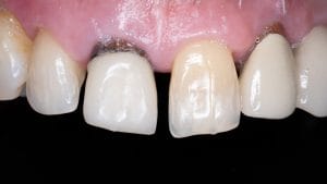
The diagnostic process for a single central crown demands particular attention. Matching one central incisor to another is arguably one of the more demanding aesthetic challenges in restorative dentistry – any discrepancy in shade, value, or character is immediately obvious. To give the laboratory the best possible information, clinical images were taken immediately on arrival at the first appointment, before dehydration could affect shade accuracy. Photographs at the correct exposure, underexposure, and greyscale were all captured and annotated before being sent to the lab alongside the intraoral scan.
Trans-sulcular probing confirmed a normal crest height of 3mm from the gingival margin – reassuring, as minimal gingival management was planned during preparation. A pre-operative polyvinyl silicone (PVS) impression was also taken at this stage as a blueprint for the temporary.
Beginning treatment
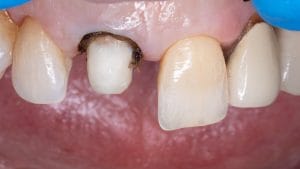
Upon sectioning and removing the existing crown, secondary caries was revealed at the margins. This was removed and the preparation extended apically towards the gingivae. The post showed no mobility on assessment and was left in situ rather than risk further root damage in attempting removal. To manage the dark underlying structure, the post was sandblasted, etched, and bonded before being masked with white opaquer followed by an A3 body shade build-up. This is a step worth emphasising: masking the post with resin rather than relying solely on the ceramic optimises subsurface scattering through the restoration and reduces the risk of a greyed gingival margin in the final result.
A two-cord retraction technique was used before an intraoral scan was taken with the Trios 3. A temporary crown in A3.5 Luxatemp was fabricated from the original PVS impression and adjusted to a good fit.
Fitting the crown
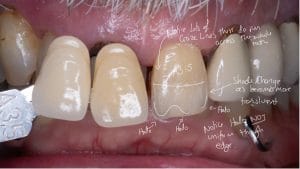
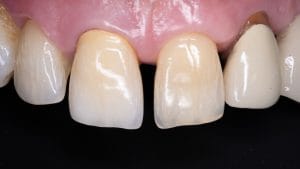
The lab’s (Byrnes) preference was monolithic zirconia; a sensible choice when it comes to masking a dark root, though value management becomes critical. At the first try-in, the value was too high against the contralateral central incisor, and the temporary had displaced the gingival margin slightly apically. Both issues were photographed and annotated, the crown was returned to the lab, and the temporary was adjusted. By the second try-in, the gingival margin had returned to its natural level and the crown’s value was considerably improved, so we agreed to cement the crown.
Following isolation with Optragate and sandblasting of the preparation, the crown was luted with RelyX Unicem dual-cure self-adhesive resin cement. Occlusion was verified with both 200µm and 20µm articulating paper to confirm no interference with the envelope of function. The patient returned one week later: the occlusion required no adjustment, gingival healing was progressing well, and he was pleased.
Reflections
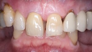
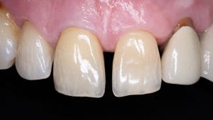
Single central crown cases are unforgiving. The contralateral tooth sets a standard that the restoration must meet in terms of shade, value, and character, and there is no hiding. In this case, I believe this to be a positive outcome, especially in the specific characteristics of the craze lines – though I will note the incisal third value is marginally lower than the natural tooth’s, and the blue halo in the contralateral central is not fully replicated. Both are observations for future cases of this type. I am grateful to the laboratory team at Byrnes for their skill and patience across multiple iterations.
For dentists approaching similar cases, two things proved decisive: thorough pre-operative documentation – including greyscale and underexposed images to guide lab character matching – and the willingness to send the crown back rather than cement a compromise. Mentorship within the BACD community has been equally important in developing the approach to complex aesthetic cases.
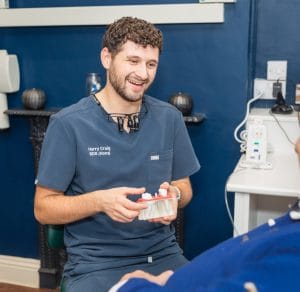
Bio: Dr Harry Craig is an Accreditation candidate for the BACD, where he also serves as Chair of the Young Membership Committee.

For more information about the BACD, please visit www.bacd.com