





Dr Minesh Patel describes a complex case in which bone augmentation using a titanium mesh was undertaken to restore a maxillary alveolar ridge with a through-and-through lesion and provide a lasting dental implant solution.
Intro
When a patient experiences a complex dental extraction, bone density may be compromised. In extreme cases, this may result in a through-and-through lesion – in which both the buccal and palatal wall are compromised. This can make treatment complex, with fewer options available for effective and long-lasting restorative solutions. Titanium mesh has helped to expand the indications of guided bone regeneration due to its mechanical properties and biocompatibility. It enables clinicians to repair alveolar ridges with large bone defects, delivering stable bone augmentation results. Traditional membranes, when applied to large defects, may be limited by their stiffness, making them difficult to maintain blood supply. Due to its good plasticity, on the other hand, titanium mesh can adapt to bony defects through bending and shaping – enabling highly stable osteogenesis and achieving bone augmentation in both horizontal and vertical directions.[i]
Patient background
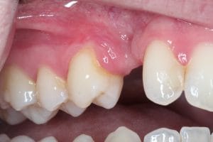
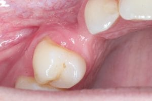
A male, 24-year-old patient presented to the practice with a missing UR3. As a result of orthodontic treatment, the patient had developed dental ankylosis – his UR3 had fused to the jaw bone. His tooth was removed at the oral surgery centre, requiring an aggressive extraction, and he was given a partial denture as a temporary solution. The patient is a professional cricketer and, due to his age, his main aim was to preserve the longevity of his dentition, with a long-term, permanent solution. Otherwise, the patient had good oral health, was compliant with treatment, but was nervous, so measures needed to be taken to manage this throughout the process.
Assessments
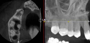
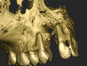
CBCT scans were taken, which revealed the extent of the buccal defect, with very minimal bone remaining at the UR3 site. This was a through-and-through lesion which would make treatment challenging, with neither a buccal or palatal wall available to provide stability.
Treatment planning
All options were discussed with the patient, taking into account the need for a long-lasting solution, the limitations due to the through-and-through lesion, and the patient’s dental anxiety. The option of doing nothing was discussed, but not recommended in this case, especially as the patient was in his early twenties. The advantages and disadvantages of options such as bridges and partial dentures were also discussed but not recommended, as they would not necessarily offer the desired long-term solution.
As such, there were two options for bone and soft tissue augmentation, followed by delayed implant placement. The option of a block bone graft was explained to the patient, which would involve harvesting bone from the lower jaw and placing it into the surgical site. However, due to the extent of the bone loss at the UR3 region, achieving blood supply would have been unreliable using this method.
The more favourable option of bone augmentation with autograft and bovine grafting material using titanium mesh was selected. It would require no donated human bone and no secondary surgical site, and this method would enable us to achieve a reliable blood supply using the surrounding tissue.
Treatment provision
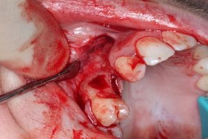
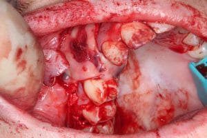
As the patient was nervous, treatment was provided under sedation, helping to keep him calm and comfortable throughout the process. A flap was raised and plasma rich membrane was harnessed to enhance tissue healing and regeneration. Bone was harvested from the patient’s palate, where a small complication occurred – bleeding wouldn’t stop at the harvest site, requiring stitching and packing to stem the bleeding. The autograft was mixed with bovine grafting material and the platelet-rich fibrin (PRF) liquid, and packed into the UR3 area. Titanium mesh was then used alongside a guided membrane technique to secure the bone grafting material, held in place with titanium tacks – two buccally, two palatally. PRF membrane was then placed over the titanium mesh to provide added coverage, accommodating the increased bone volume. Prolene 5-0 sutures were excellent for securing this, and good gingival thickness was achieved. CBCT imaging showed that the fullness of the bone had been restored.
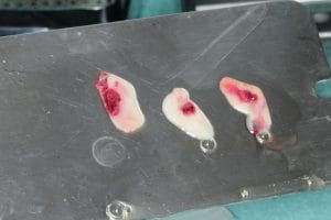
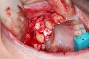
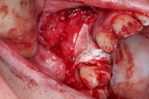
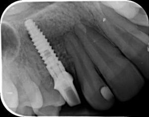
The site was left to heal for 6 months, during which time the patient was provided with a carbon fibre temporary restoration. A BioHorizons Tapered Internal 3.8 x 15 mm implant on a 3.5 platform was placed. This implant was selected as it required minimal drilling and allowed for good primary fixation. Axion Dental Lab provided a custom abutment cast in zirconia to improve aesthetics, which was a concern due to the region it was in. Occlusion was adjusted to establish group function and distribute occlusal forces rather than relying on canine guidance – therefore reducing the impact on the new implant.
Outcome and case appraisal
My patient and I were extremely happy with the treatment result. Due to his age, it was important to create a long-lasting outcome.
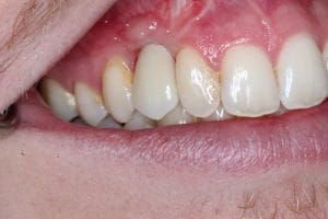
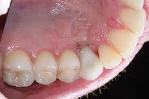
There were a number of factors that contributed to this being a challenging case: the surgical site was located in the smile line, there was a severe lack of bone, and we wanted to achieve a good gingival margin, implant angle, and occlusion for a functional and aesthetic outcome. Additionally, the patient was required to be engaged in a lengthy treatment, meaning maintaining compliance was a potential obstacle.
This was also a complex and technically difficult case, requiring the implant to be placed solely in grafted bone. As such, it required very high-quality bone and soft tissue augmentation. Usually, when providing a graft using a titanium mesh, there is one wall to work with – in this case neither the buccal or palatal wall was available.
Further to this, handling titanium mesh is very difficult. It’s important that clinicians undergo mentoring and training prior to attempting this, because it is tactically more difficult to place pins, and practitioners must be comfortable with raising a palatal flap.
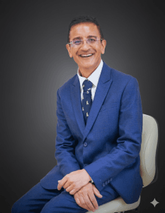 Author Bio: Dr Minesh Patel qualified from Cardiff in 1986 and has been placing dental implants for 32 years. He gained his MFGDP in 1996, the Diploma in Implant Dentistry in 2005, his MSc in Implantology in 2006, and his MSc in Restorative and Aesthetic Dentistry in 2016 from Manchester University. Dr Patel holds a primary qualification in Dental Law and a PG Cert in Education both from Bedfordshire University. He is also an accredited Mentor for the Association of Dental Implantology. Dr Patel has been awarded as course director for Implantology at Warwick University, and is the Surgical Module Lead for block grafting, sinus grafting and soft tissue grafting for the Masters students. Dr Patel is an examiner for the Diploma in Implant Dentistry at the Royal College of Surgeons of England.

For product information or further education from BioHorizons and Camlog, please visit https://theimplanthub.com/
[i] Xie, Y., Li, S., Zhang, T. et al. Titanium mesh for bone augmentation in oral implantology: current application and progress. Int J Oral Sci 12, 37 (2020). https://doi.org/10.1038/s41368-020-00107-z


