





CBCT, intraoral scanning, digital treatment planning, artificial intelligence – talk on dental imaging is currently centred around the new. And rightly so. These technologies are all actively changing what is possible when it comes to the dental practice.
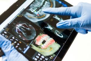
But there is another argument worth making alongside all of that, and it is this: the intraoral radiograph remains the most frequently taken diagnostic image in general dentistry by a considerable distance. Periapical and bitewing radiographs are taken in virtually every practice, on virtually every patient, at virtually every relevant appointment. The treatment decisions that follow from them – whether caries is present, whether it has reached the pulp, whether bone levels are changing – are among the most crucial a GDP might make in the course of a normal working week.
And if that is true, then the quality of the sensor used to take those images deserves at least as much attention as the more headline-grabbing end of the imaging spectrum.
Good radiography is a powerful tool
The resolution and image quality of intraoral radiography has consequences that are direct and highly important. Interproximal decay at an early stage can be invisible on a lower-resolution image yet detectable on a higher-resolution one. The difference between catching a lesion at that stage and catching it six months later, when it has progressed, can mean the difference between preventive advice and a sizeable restoration.[i]
Similarly, to properly diagnose periapical issues in the early stages requires fine detail, which is something that the sensor quality directly determines. A clinician working with a high-resolution sensor, and the ideal image processing software for the job, is the one with a more diagnostically useful radiograph when it comes down to it.
Dose and the justification principle
The case for high-quality intraoral sensors is also seen as an ionising radiation (medical exposure) regulations (IRMER) argument. The justification and optimisation principles that are in charge of dental radiography in the UK require that every exposure is justified by the benefit it will provide, and that doses are kept as low as reasonably achievable.[ii] A sensor that can achieve diagnosable images at a lower dose will directly support that obligation.
Dose optimisation can be framed as a secondary consideration on occasion; i.e., something to address once the quality for diagnosis has been established. A better way to frame this is that sensor quality and dose efficiency are the same conversation. Since a sensor with enough resolution to produce useful images at reduced exposure settings is delivering it more efficiently.
Consistency and the team
In a practice with multiple team members taking radiographs, the consistency of images is an important consideration to address. Variable image quality across team members, as a result of factors such as differences in technique, creates variability in the diagnostic information available at review. Standardised, high-quality sensors with reliable image output across operators are crucial for reducing variability and therefore improve reproducibility and diagnostic capability.
This is particularly relevant when monitoring progressive conditions where the ability to compare images taken at different appointments at different times is vital, and based entirely on consistent image quality.
Integration and workflow
Sensor investment extends easily to workflow. If a sensor can integrate simply with the already existing imaging software, allowing images to be captured and viewed without file transfer or format conversion, it saves time and reduces the risk of images being lost or difficult to find. This is not a small consideration when it comes to busy practices.
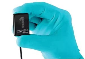 The Carestream Dental RVG 6200 is a high-resolution intraoral sensor designed for the kind of everyday diagnostic work that is integral to general practice. With a pixel pitch of 19.5 microns and a sensor size available in both size 1 and size 2, the RVG 6200 delivers sharp, diagnostically rich images at optimised dose levels, with seamless integration into CS Imaging 8 and compatibility with a wide range of third-party software. Its rounded corners and flexible cable are designed for patient comfort as well as for ease of use across long lists and busy clinics.
The technology set to change what is possible in dentistry is having its place, understandably, in the spotlight right now. It’s time to revisit the technology that makes everyday diagnosis more reliable. For many practices, these may not be the same investment – but both are invaluable.

For more information on Carestream Dental visit www.carestreamdental.co.uk
For the latest news and updates, follow us on Facebook and Instagram @carestreamdental.uk
[i] Clarkson JE, Pitts NB, Goulao B, et al. Risk-based, 6-monthly and 24-monthly dental check-ups for adults: the INTERVAL three-arm RCT. Southampton (UK): NIHR Journals Library; 2020 Nov. (Health Technology Assessment, No. 24.60.) Chapter 1, Introduction. Available from: https://www.ncbi.nlm.nih.gov/books/NBK564217/
[ii] Department of Health and Social Care. Ionising Radiation (Medical Exposure) Regulations 2017: guidance. GOV.UK. Published 2017. https://www.gov.uk/government/publications/ionising-radiation-medical-exposure-regulations-2017-guidance


