





Dr Alex Payne restores a dentally anxious patient’s lower dentition with same day results
Introduction
A considerable proportion (over 15%) of the global population have dental fear and anxiety.[i] This is a multifactorial issue that impacts individual’s access to dental care and, therefore, their oral health.[ii] It is vital for professionals to acknowledge and manage dental fear to ensure that patients can complete the treatment they need. The literature suggests several strategies to consider, from distraction therapy to hypnosis, acupuncture, and virtual reality.[iii]
In some situations, dental anxiety can be improved with clear communication from the practitioner and fewer treatment steps. Immediate implantology offers a solution, enabling restoration of oral function and aesthetics in a single surgery, when appropriate. The below case details a patient’s preference for immediacy to help overcome her dental fear.
Patient presentation
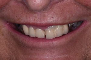
A 60-year-old female patient presented with significant dental anxiety and treatment phobia. Her chief complaint was the repeated debonding of an existing upper left bridge in the UL1–3 region. She reported that the bridge frequently became dislodged and that she had been re-cementing it daily using over-the-counter temporary dental cement. The situation was causing significant frustration and functional difficulty.
The patient was also aware that several other teeth had previously fractured or required extraction and expressed concern regarding the long-term prognosis of her remaining dentition.
Clinical examination and diagnosis
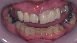
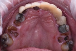
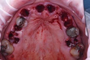
Following removal of the existing bridge, the retained roots beneath the abutments were found to be unrestorable due to extensive caries extending subgingivally. Additional unrestorable retained roots were present at UR5, UR6 and UL6.
Among the remaining teeth, UL5 and UR4 were heavily restored with large amalgam restorations and exhibited associated periapical pathology.
At this stage the only maxillary teeth considered to have a favourable long-term prognosis were UR1, UR3, UR7 and UL7, although the condition of UR1 and UR3 remained uncertain due to the previously existing bridge.
Treatment planning
A detailed discussion was undertaken with the patient regarding possible treatment options. It may have been possible to retain any remaining teeth with a favourable prognosis and place implants between them to restore the missing sites. The alternative was extraction of the remaining maxillary teeth and rehabilitation with a full-arch implant-supported restoration.
Due to the patient’s significant dental anxiety, she expressed a preference to complete as much treatment as possible in a single stage. A full-arch implant solution was therefore selected.
The prosthetic design was discussed in relation to FP1 and FP3 restorative concepts. As the patient presented with favourable bone levels, good existing arch architecture and a thick soft tissue biotype, an FP1 restoration was considered appropriate. The patient’s tooth loss was not associated with poor oral hygiene, and therefore the proposed FP1 restoration was considered maintainable long-term.
Restorative treatment was also planned for the mandibular arch to improve occlusion and aesthetics.
Digital planning and guided surgery
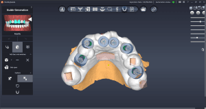
Planning was performed using a facially driven smile design. Once the proposed aesthetic outcome had been agreed with the patient, a digital wax-up was created and combined with CBCT data in the planning software Dentiq. This allowed prosthetically driven implant placement to be planned.
Two surgical guides were fabricated in-house using a Phrozen Sonic Mini 8K S printer and NextDent SG resin.
The first guide was tooth-supported and would be used to place six implants while the remaining teeth were still present. The remaining teeth would then be extracted and a second guide used to complete implant placement.
This second guide would be implant-supported using the CONELOG® Progressive screw carriers (Camlog), allowing accurate guide stabilisation without the need for bone fixation pins or reliance on soft tissue support.
Implant selection
CONELOG® Progressive-Line implants (Camlog) were selected due to their predictable primary stability, which is important for immediate loading protocols. The conical implant-abutment connection provides a stable restorative interface, and the COMFOUR multi-unit abutment system (Camlog) offers a slim emergence profile that is well suited to FP1 restorations.
This design allows implants to be placed slightly subcrestally while maintaining favourable prosthetic contours. In posterior regions, the larger 5.0 multi-unit abutments provide an improved emergence profile without the need to position implants excessively deep.
The SLA surface and grade 4 titanium composition also provide reliable and predictable osseointegration.
Surgical procedure
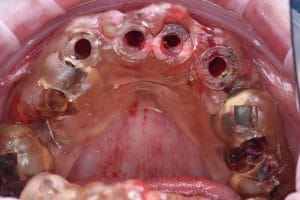
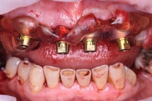
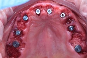
A total of eight implants were placed in positions corresponding to those shown in the clinical imaging. All implants were placed in a prosthetically guided manner so that straight multi-unit abutments could be used.
All implants achieved insertion torque values exceeding 35 Ncm, allowing immediate loading.
MinerOss Putty (BioHorizons) was used to graft the residual gaps and preserve the pontic sites. This material was selected due to its ease of handling and its collagen-containing allograft composition, which provides structural support during healing while gradually resorbing.
Although soft tissue grafting is commonly performed in full-arch immediate cases, this patient presented with favourable tissue architecture and a thick biotype. For this reason, additional soft tissue grafting was not performed.
Immediate provisionalisation
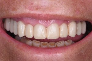
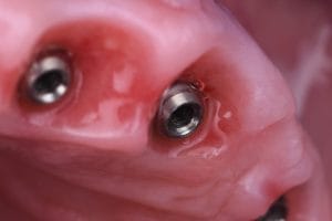
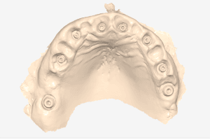
Following surgery, an intraoral scan was taken using a Medit i700 scanner with PEEK scan bodies.
Prior to surgery, the brilliant Smile Design Laboratory had already designed the majority of the provisional restoration based on the digital wax-up. Using the post-operative scan data, the FP1 provisional restoration was finalised and milled in graphene.
The provisional bridge was delivered a few hours after surgery and fixed directly to the multi-unit abutments using Rosen screws. The restoration was screw-retained and demonstrated a passive fit on first insertion.
Healing and review
At the one-week review appointment, soft tissue healing was excellent and the patient reported minimal post-operative discomfort.
Healing progressed uneventfully and at three months the soft tissue architecture was stable with well-integrated peri-implant tissues. The patient was very satisfied with the aesthetic and functional outcome.
During the healing period, the planned restorative treatment in the mandibular arch was also completed.
Final restoration
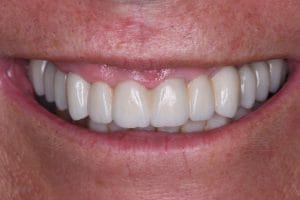
A further digital scan was taken with the Medit i700 to fabricate the final prosthesis. Particular care was taken to scan the fitting surface of the provisional restoration so that the established contours and emergence profile could be replicated.
The definitive restoration was delivered two weeks later. It consisted of a monolithic zirconia FP1 prosthesis characterised with staining and glazing and supported on TiBases.
Both the provisional and final restorations were screw-retained and demonstrated a passive fit on first insertion. Ceramic surfaces in contact with the peri-implant soft tissues were left unglazed and highly polished to support soft tissue health.
Outcome
At delivery, the patient was delighted with the final result. Clinical review demonstrated excellent soft tissue integration and stable peri-implant tissues.
This case demonstrates how a digitally guided and prosthetically driven workflow can be used to manage a terminal maxillary dentition efficiently. Guided implant placement combined with immediate loading allowed a predictable full-arch FP1 rehabilitation while minimising the number of treatment stages for a highly anxious patient.

For product information from BioHorizons and Camlog, please visit https://theimplanthub.com/
 Author bio: Dr Alex Payne practises at Greasby Dental Centre in the Wirral. He has completed an Implant Diploma with the Royal College of Surgeons and is the Clinical Lead of the Smilefast Implant Diploma.
[i] Silveira ER, Cademartori MG, Schuch HS, Armfield JA, Demarco FF. Estimated prevalence of dental fear in adults: A systematic review and meta-analysis. J Dent. 2021 May;108:103632. doi: 10.1016/j.jdent.2021.103632. Epub 2021 Mar 9. PMID: 33711405.
[ii] Piechal, Agnieszka & Siekierska, Edyta & Blecharz-Klin, Kamilla. (2025). Etiology of Dental Anxiety and Dental Phobia: Review. European Journal of Dentistry. 20. 10.1055/s-0045-1809146.
[iii] Hoffmann B, Erwood K, Ncomanzi S, Fischer V, O’Brien D, Lee A. Management strategies for adult patients with dental anxiety in the dental clinic: a systematic review. Aust Dent J. 2022 Mar;67 Suppl 1(Suppl 1):S3-S13. doi: 10.1111/adj.12926. Epub 2022 Jul 12. PMID: 35735746; PMCID: PMC9796536.


