





Dr Ravinder Jhutie describes a case in which immediate dental implant treatment was carried out to restore an upper anterior incisor.
Intro
The immediate placement and loading of dental implants in the provision of single maxillary anterior restorations is considered a predictable and safe treatment option, resulting in adequate survival rates and favourable aesthetic outcomes for five years post-surgery.[i] Further contributing to high survival rates is the use of guided implant placement and implant planning software, with research suggesting that a guided approach is less invasive than a standard protocol, requiring less time and causing minimal discomfort. The use of digital planning software is also advantageous, making it possible to plan the position of implants prior to surgery to a high degree of accuracy. This also allows clinicians to anticipate the potential changes or complications that might occur and mitigate these ahead of time.[ii]
Patient background
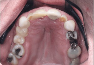
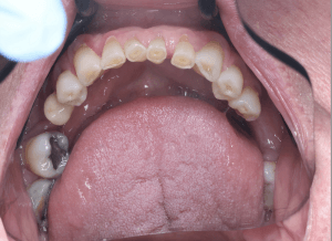
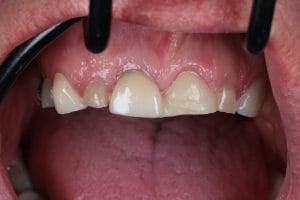
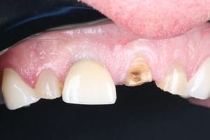
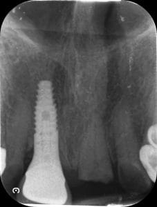
A 68-year-old male patient attended the practice with failure of the UL1. He had a temporary restoration provided by his regular dentist, who then referred him for a permanent solution. Overall, the patient’s oral health was good, with a number of existing restorations in the mouth, including an implant at the UR1 (present for 10+ years), and considerable tooth wear present throughout the dentition. The patient was not interested in full-mouth rehabilitation – his primary goal was to restore the function and aesthetics in the anterior region, with a fixed replacement for the UL1.
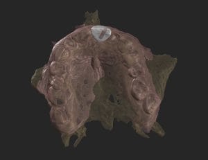
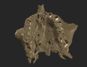
A range of treatment options were discussed with the patient, including dentures, bridges, and dental implants, ensuring he understood the risks and benefits of each. Ultimately, the patient chose a dental implant as he preferred to have a fixed, gold-standard solution. A comprehensive assessment was then carried out to ensure the patient was a suitable candidate for dental implant treatment, including a 3D scan to assess the bone and the patient’s anatomy, and intraoral scans to facilitate digital planning.
Treatment planning
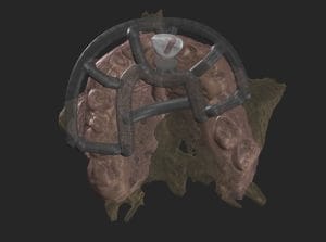
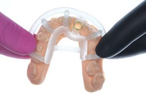
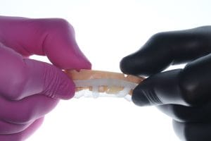
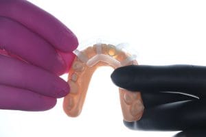
In this case, a guided surgery was preferred in order to facilitate a prosthetically-driven approach. This would ensure that the positioning and angle of the implant was correct, and would create the ideal aesthetic and functional outcome required in the anterior zone.
Additionally, an immediate placement and loading approach was selected for this case. Particular care was required with the mesial and distal soft tissue during removal of the temporary restoration and extraction of the root – with the aim to keep the surrounding soft tissue intact.
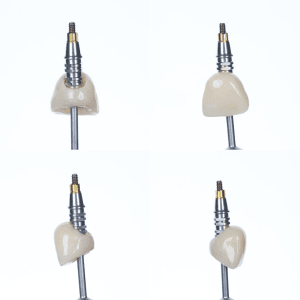
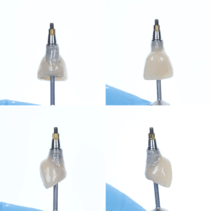
A CBCT and intraoral scans were taken and uploaded to SMOP as well as Smilecloud software for digital planning alongside the dental technician (Sohaib Abouelela at Zenith Dental Labs). This planning stage included designing the surgical guide as well as a digitally-produced crown. This software allows dental professionals to change the shape of the tooth, enabling the crown to be designed in such a way that the soft tissue graft could grow and expand in an ideal way. The surgical guide was fabricated prior to the appointment in the dental lab, as was the crown, which was then attached to the temporary abutments with composite to create the ideal emergence profile, as well as aid with pink aesthetics.
Treatment provision
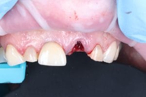
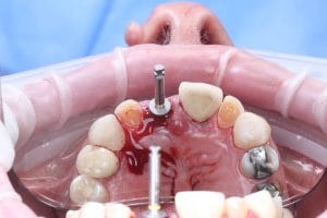
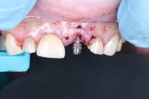
On the day of surgery, the temporary restoration was removed, and the UL1 root was extracted as atraumatically as possible, with the mesial and distal soft tissue kept intact according to the clinical plan. This ensured that there was a good blood supply between the palatal and buccal area, preserving the health of the bone and soft tissue.
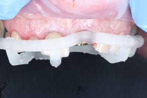
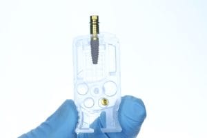
The surgical guide was placed in the mouth, and the site was drilled in accordance with the surgical plan. One screw-mounted 3.8mm x 11mm CONELOG® Progressive-Line Promote plus implant (Camlog) was placed through the guide, following the treatment plan with precision.
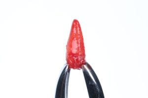
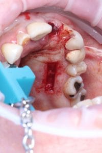
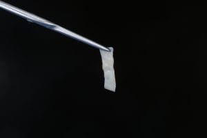
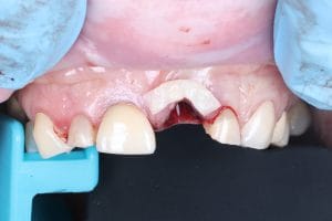
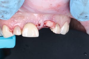
To ensure sufficient soft tissue volume could be achieved at the UL1 site, a free gingival graft was harvested from the palate and de-epithelialised to obtain connective tissue. The soft tissue graft was placed, and stabilised with three sutures (Glycolon 5.0).
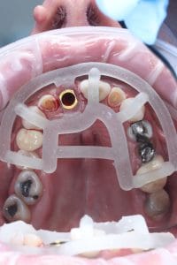
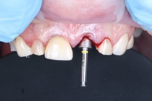
A temporary cylinder abutment was then placed, and bone grafting was carried out using MinerOss® Blend (BioHorizons) – consisting of 70% cortical and 30% cancellous allograft chips. This aimed to fill the jump gap between bone and implant; this was carefully placed in the bone corridor to aid with reliable revascularisation to improve bone volume at the site, and aid stable healing.
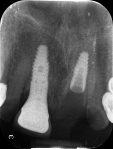
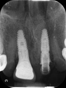
Following this, the restorative guide was used to place the crown into the mouth. This is done to ensure that the resulting prosthesis will be seated in the correct 3D position, mesially, distally, buccally, and palatally, as was planned on the digital smile design. Once this was confirmed to be correct, the implant was restored out of occlusion with the provisional restoration, and a post-operative radiograph was taken to ensure that the implant and crown were seated correctly.
Outcome and case appraisal
The case went extremely well. The patient and I were very happy with the outcome. The patient was not in pain, and at four days post-op, the site was healing really well.
This was my first time using the CONELOG® Progressive-Line Promote plus implant, and I was very impressed with its performance. It is a system which my mentor, Dr Imran Nasser uses, and recommended to me. It is a versatile option for immediate placement and loading, especially as it offered excellent torque values.
Utilising a digital planning system made it really easy to liaise with the dental technician in this case, meaning that we were able to really effectively plan the whole case, including finalising implant diameter and length, ensuring that as much possible has been planned for prior to the day of surgery.
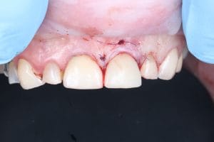
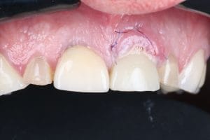
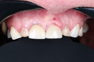
My advice for clinicians who are planning to undertake a similar case would be to take your time – for example, allocating more time than you think you’ll need, booking four hours for what could be a one-hour surgery, removing time pressure and allowing you to give your full focus to the patient in your chair. I personally also find it helpful to write out the treatment steps, so that I feel confident that the treatment plan is being followed with complete accuracy. This systematic approach, along with tools such as digital planning and guided surgery, has had a big impact for me, and has made the results I can achieve consistent and repeatable.

For product information from BioHorizons and Camlog, please visit https://theimplanthub.com/
 Author bio: Dr Ravinder Jhutie graduated from the University of Birmingham in 2017, and provides a range of treatments, including dental implants. Dr Jhutie has a passion for creating life-changing smiles for his patients and, in 2022, acquired TopLabs dental laboratory to ensure the quality and precision of the dental prosthetics he provides matches that of his surgical capabilities. He has now sold Toplabs prosthetics and ventured into other industries including The Haulage Family. Dr Jhutie has a wide collection of interests and, as part of his self-development, believes there is value in being uncomfortable to achieve progress.
[i] Wittneben, J.G., Molinero‐Mourelle, P., Hamilton, A., Alnasser, M., Obermaier, B., Morton, D., Gallucci, G.O. and Wismeijer, D., 2023. Clinical performance of immediately placed and immediately loaded single implants in the esthetic zone: a systematic review and meta‐analysis. Clinical Oral Implants Research, 34, pp.266-303.
[ii] Dioguardi, M., Spirito, F., Quarta, C., Sovereto, D., Basile, E., Ballini, A., Caloro, G.A., Troiano, G., Lo Muzio, L. and Mastrangelo, F., 2023. Guided dental implant surgery: Systematic review. Journal of clinical medicine, 12(4), p.1490.


