


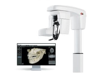
3D imaging in dentistry, now accessible for many dentists through cone beam computed tomography (CBCT) scanners, has created new insights into patients’ oral health. This allows clinicians to better inform their treatment plans, especially in cases requiring surgical intervention, such as implant placement or tooth extractions.
In order to make effective decisions which lead to predictable treatment outcomes, dental professionals need a clear view of the dentition.
Dental professionals need to be aware of challenges that may be present, and how they can affect the quality of a radiographic image. For example, the targeted area may be obscured, or key physiological structures may be omitted, necessitating a second CBCT assessment.
Metal artefact presence
A CBCT scan can be affected in a number of ways, with artefacts (streaking, shadowing, ringing and cupping) misrepresenting the actual dentition of a patient.[i] These can have a number of potential causes, and one of the most notable for dental professionals is metal restorations. This includes previous restorative solutions made from materials such as amalgam or gold alloy, and can also include aspects such as dental implants.
A metal restoration absorbs lower wavelength X-rays, creating a ‘hardening’ effect.[ii] This influences the image quality by reducing contrast, obscuring structures, and making it difficult to suitably analyse nearby structures. This can make diagnosis difficult and time-consuming;ii in some cases, a repeated CBCT scan may be required, resulting in additional exposure to X-ray radiation for a patient. CBCT systems typically produce a higher radiation dose than conventional dental radiography equipment,[iii] and repeated exposures should be aimed to be minimised where at all possible, following the ALARA (As Low As Reasonably Achievable) principle.[iv] Whilst radiation from a single exposure is as low as 12-30 days of normal background radiation,[v] repeat scans increase the risk of adverse outcomes over time.
The difficulty faced by clinicians is the sheer prevalence of metals in restorative dental care. Alongside their use in dental implants or post crowns, the British Dental Association notes that amalgam could be used for around a third of all fillings placed in England on the NHS, though there are no official figures to confirm this.[vi]
To avoid metal artefacts, clinicians can try to evade the affected area entirely by minimising the field of view. This is not always possible, and so some of the latest CBCT solutions find new ways to mitigate the effects through metal artefact reduction algorithms. These help to recreate an image of the dentition that is affected by hardening, with effective options allowing a comparison between results with and without the effect, ensuring the most accurate diagnosis can be achieved and misinterpretation is minimised.
Patient movement
CBCT systems have an acquisition time ranging anywhere from 5.4 to 40 seconds.[vii] This creates the potential for patients to move during the scan, disrupting the process and causing motion artefacts. Whilst patients are reminded to keep still during a scan, movement may be entirely unconscious, and the result is a scan with decreased quality, demanding a retake.
The literature notes that claustrophobia, being very young or very old, and a fear of the CBCT scan can all be potential causes of patient movement.[viii] Anxiety in particular can create both an emotional and physical response, causing feelings of tension and jumpiness, restlessness, dizziness, shortness of breath, and tremors and twitches.viii
Traditional approaches to patient management in the case of dental anxiety may be helpful. This includes explaining the CBCT procedure in language that the patient will understand whilst listening to and responding to concerns. It’s important to explain what patients can expect, including how long the scan will take, and how long they need to keep still for, as well as preparing them for any moving parts or sounds that will occur. Keeping patients comfortable is key, and a versatile scanner will be able to aid patients who want to complete the scan standing or seated.
Choose your solution
 Whilst it may feel as though artefacts make an impact that is often outside the clinician’s control, the choice of CBCT system can make a significant difference.
The CS 8200 3D Advance Edition CBCT system from Carestream Dental features Metal Artefact Reduction technology, which can aid confident diagnoses by eliminating some of the effects of metal restorations. Clinicians can immediately compare with an untouched scan to ensure accuracy at every step. Plus, the CS 8200 3D Advance allows for standing and seated scans, with a comfortable scan zone for patients.
Dental professionals can expect artefacts as an inevitability in dental radiography. What is important is how they aim to minimise such occurrences, and how patients are cared for if a retake is needed. This is all only possible with high-quality equipment, and complete clinical knowledge.
For more information on Carestream Dental visit www.carestreamdental.co.uk
For the latest news and updates, follow us on Facebook and Instagram @carestreamdental.uk
 Author: Nimisha Nariapara – Trade Marketing Manager at Carestream Dental covering the UK, Middle East, Nordics, South Africa, Russia and CIS regions.
[i] Xie, S., Liang, Y., Yang, T., & Song, Z. (2020). Contextual loss based artifact removal method on CBCT image. Journal of Applied Clinical Medical Physics, 21(12), 166-177.
[ii] Omar, G., Abdelsalam, Z., & Hamed, W. (2016). Quantitative analysis of metallic artifacts caused by dental metallic restorations: comparison between four CBCT scanners. Future Dental Journal, 2(1), 15-21.
[iii] HPA Working Party on Dental Cone Beam CT Equipment. (2010). Guidance on the Safe Use of Dental Cone Beam CT (Computed Tomography) Equipment. (Online) Available at: https://assets.publishing.service.gov.uk/media/5a7d4f13ed915d321c2de451/HPA-CRCE-010_for_website.pdf [Accessed November 2025]
[iv] Habibi, Y., Habibi, E., & Al-Nawas, B. (2019). Re-exposure in cone beam CT of the dentomaxillofacial region: a retrospective study. Dentomaxillofacial Radiology, 48(3), 20180184.
[v] NHS Calderdale and Huddersfield NHS Foundation Trust, (n.d.). Cone Beam CT (CBCT). (Online) Available at: https://plr.cht.nhs.uk/download/1420/Cone%20Beam%20CT%20%28CBCT%29%20A4 [Accessed November 2025]
[vi] British Dental Association, (2024). Government Failure on amalgam ban could break NHS dentistry. (Online) Available at: https://www.bda.org/media-centre/government-failure-on-amalgam-ban-could-break-nhs-dentistry/ [Accessed November 2025]
[vii] Hatcher, D. C. (2010). Operational principles for cone-beam computed tomography. The Journal of the american dental association, 141, 3S-6S.
[viii] Yıldızer Keriş, E. (2017). Effect of patient anxiety on image motion artefacts in CBCT. BMC oral health, 17(1), 73.


