





Dr Imran Nasser presents an aesthetically-challenging immediate implant case.
Introduction
Immediate implant placement has been proven successful in various areas of the maxilla and mandible, including the canine sites.[i] However, treatment in these regions can present specific challenges that clinicians have to be prepared for in order to ensure treatment success. The digital workflow affords one possible solution, with the literature showing this approach to increase accuracy of implant placement in a range of clinical situations.[ii]
In the presentation that follows, the author explores the use of an immediate implant in an aesthetically-demanding case. Emphasis was placed on meticulous digital planning and appropriate implant selection to minimise the potential risks and complications involved.
Case presentation
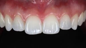
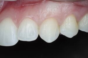
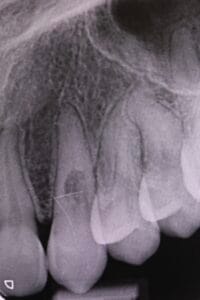
A 25-year-old female patient was referred due to internal resorption on the UL3. She had previously undergone orthodontics and was generally happy with her smile, though smile aesthetics was a priority.
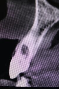
A comprehensive clinical assessment was conducted to determine treatment options for the UL3. The patient’s oral health was good with an otherwise unrestored mouth. A high smile line was recorded, which would be important to address for the aesthetic outcome. A CBCT scan revealed extensive resorption, deeming the UL3 unrestorable. This was explained to the patient, discussing all potential treatment options from doing nothing to a bridge and an implant-retained crown. The latter was preferred by the patient.
Treatment planning
Planning was key in this case given that the patient had such high aesthetic expectations. The clinical photos, intraoral scans and a CBCT scan were integrated to facilitate digital planning, which was conducted on Smilecloud software.
A thin biotype, combined with proclined incisors – meant that reduced bone thickness was present on the facial aspect of the teeth. As such, the implant position was crucial – it needed to be placed as palatally as possible, with a large jump gap for maximum grafting material, to ensure an aesthetic and functional result, as well as a decreased risk of implant thread exposure.
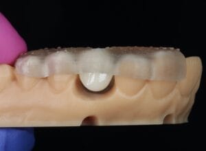
Steps were also taken to ensure that the crown would be placed without functional load – this is always more challenging when operating at the canine site. However, upon reflection of all the diagnostic data, the author was confident that this could be achieved in this case. The digital plan also meant that the anatomy of the canine could be better visualised, allowing full duplication and the creation of a custom healer. This component provided a contingency option in case the canine couldn’t be immediately loaded on the day of surgery.
The digital prosthetic plan was transferred to SMOP software to determine the appropriate implant position, angle, and depth. The SMOP digital guide was fabricated in anticipation of surgery. The entire clinical procedure was explained to the patient in detail and informed consent was obtained.
Surgery
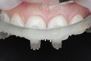
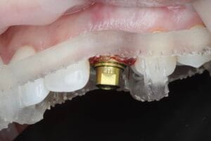
Another challenge when attempting immediate loading on a canine implant is the atraumatic extraction of the failing tooth while maintaining sufficient buccal plate. In this case, the tooth was removed completely, without sectioning, using forceps.
The SMOP guide was then used to place the implant. Osteotomy preparation followed the typical CONELOG® drilling sequence and a 3.8mm x 13mm CONELOG® Progressive-Line implant (Camlog) was placed in the predetermined location. The length was selected to ensure a minimum of four implant threads were placed in bone for optimal primary stability – this implant was torqued to 65Ncm. The width was also ideal for ensuring a jump gap of at least 2.5mm between the implant and buccal bone. With regards to depth, the implant was placed 1mm below the buccal plate and 4mm below the level of the predicted CEJ, respecting biological principles for enhanced outcomes.
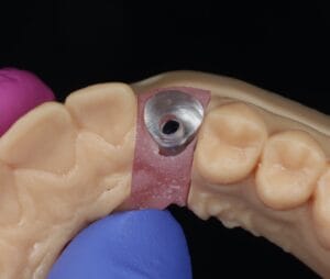
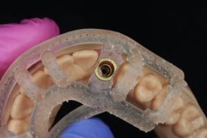
A temporary cylinder was used to make the temporary restoration. A custom jig was fitted to hold the provisional crown and to pick it up for a try-in. Outside of the mouth, composite was added to improve the emergence profile, before polishing the crown with silicone rubbers.
Both bony and soft tissue remodelling occurs naturally following extraction.[iii] To minimise the amount of resorption and recession that would happen, and to improve soft tissue outcomes, a connective tissue graft was performed. Donor tissue was harvested from the palate and sutured into place, positioned from papilla to papilla. This was placed 1mm below the free gingival margin on the internal aspect.
MinerOss® Blend allograft (BioHorizons) was packed into the jump gap around the implant. Combining cortical and cancellous bone particles, I find this material delivers a fast turnover of bone, with good density and predictable revascularisation.
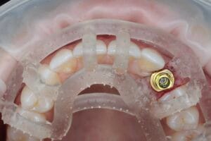
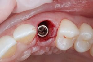
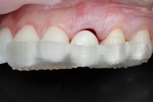
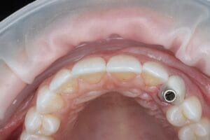
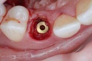
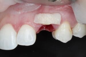
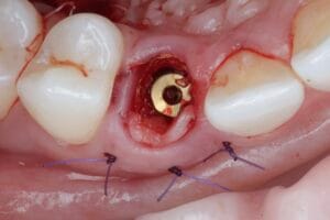
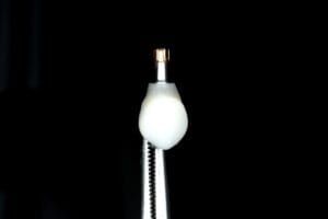
The provisional crown was then seated, the occlusion was checked, and the restoration adjusted to ensure that it was out of function. This eliminated the need for the pre-fabricated customer healer, but this is always an excellent fallback approach.
Upon conclusion of treatment, the patient was sent away with standard post-operative instructions to support healing and recovery.
Outcome and reflections
Upon review 6-months post-operatively, the patient reported the provisional to be very comfortable and to be happy with its aesthetics. The final restoration will be provided shortly, but the slight delay was important for the patient’s budget.
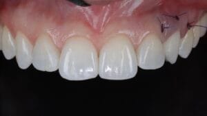
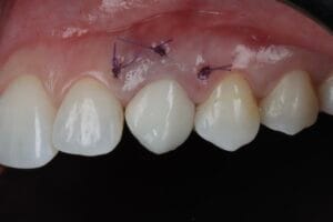
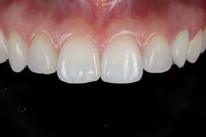
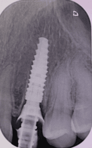
This patient displayed all the risk factors for aesthetic complications. Grafting was vital to minimise these risks. The CONELOG® Progressive-Line implant is excellent for immediate situations due to the tapered design and side-cutting preparation drills leading to high primary stability. The positive seating of the restoration into the internal connection and the tight hermetic seal provide confidence in immediate situations.

For product information from BioHorizons and Camlog, please visit https://theimplanthub.com/
 Author bio: Dr Imran Nasser is Practice Principal at Cheltenham & Cotswold Dental. He completed his MFDS in 2009 and his Masters degree in Implantology in 2014. He runs regular courses in the UK for implant & ceramic restoration (Aesthetic Prosthetic), Ridge Preservation (15C) and Immediate placement & Soft tissue grafting (Replace). For the past four years in succession (2021-2024), Imi has received the accolade of winning the UK Aesthetic Dentistry & Clinical Case Awards in Implant & Ceramic categories. He is passionate about sharing his experiences with his colleagues (www.mxicourses.com).
[i] Ickroth A, Christiaens V, Pitman J, Cosyn J. A Systematic Review on Immediate Implant Placement in Intact Versus Non-Intact Alveolar Sockets. Journal of Clinical Medicine. 2025; 14(7):2462. https://doi.org/10.3390/jcm14072462
[ii] Abad-Coronel C, Vandeweghe S, Vela Cervantes MD, Tobar Lara MJ, Mena Córdova N, Aliaga P. Accuracy of Implant Placement Using Digital Prosthetically-Derived Surgical Guides: A Systematic Review. Applied Sciences. 2024; 14(16):7422. https://doi.org/10.3390/app14167422
[iii] Tan, Wah & Wong, Terry & Wong, May & Lang, Niklaus. (2012). A systematic review of post-extractional alveolar bone dimensional changes in humans. Clinical oral implants research. 23 Suppl 5. 1-21. 10.1111/j.1600-0501.2011.02375.x.


