







Zygomatic implants are a valuable solution for patients with severe maxillary atrophy who are unsuitable for conventional implants or bone grafting. Anchored in the zygomatic bone, these implants bypass the need for vertical bone height in the posterior maxilla.
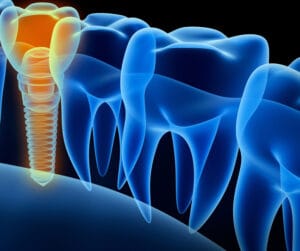
While they offer high success rates and life-changing results, they also come with distinct risks and complications that clinicians must be prepared to address or refer if necessary.
Surgical complexity and anatomical risks
Placement demands precise anatomical knowledge and surgical skill. Proximity to the orbit, infraorbital nerve, and maxillary sinus increases intraoperative risk. Misplacement may cause orbital penetration, nerve damage, or sinus perforation.[i]
Sinus complications
Sinusitis is one of the most frequently reported complications.[ii]
Sinus perforation during zygomatic implant placement typically occurs due to anatomical challenges brought about by its proximity to the surgical site. The thinness and variability of the sinus wall and Schneiderian membrane make it prone to accidental perforation. [iii]
This is particularly problematic given that patients who suffer from atrophic maxilla will typically present with a thinner wall between the sinus floor and the alveolar crest, thus the accidental breaching of the Schneiderian membrane is more likely.
Chronic sinus infections caused by sinus perforation compromise implant stability and cause significant discomfort for the patient. ii Some of the long-term complications include on-going nasal congestion, nasal discharge, facial pain/pressure, headaches, and a reduced sense of smell.[iv]
For these reasons, it is vital that clinicians are able to thoroughly analyse the anatomical characteristics of each patient using advanced imaging technologies.
Soft tissue issues
Due to the angulated emergence of zygomatic implants, soft tissue management is critical. Poor emergence profiles can lead to mucosal irritation and peri-implantitis.v
A prosthetic design which is overly bulky can cause the patient significant difficulties in their ability to maintain adequate oral hygiene, leading to the accumulation of bacterial biofilm around the implant. Controlling this is crucial to prevent the onset of peri-implantitis.[v]
Inadequate keratinised mucosa around the implant may also contribute to long-term complications. Some studies suggest that healthy keratinised mucosa may protect the surgical site against microorganisms and subgingival plaque, thus decreasing the likelihood of peri-implantitis.[vi]
To allow for the patient to carry out adequate home-hygiene, clinicians should avoid over-contoured prosthetics that trap food and plaque, opting for smooth transitions between implant and soft tissue to reduce bacterial niches.v
Prosthetic challenges
Restorative planning for zygomatic implants can be complex. Achieving proper occlusion, aesthetics, and function requires careful coordination between surgical and prosthetic teams.[vii]
Poor implant positioning can also contribute to serious complications including orbital and ocular penetration[viii] or damage to the infraorbital nerve during the surgical drilling process due to improper preparation and lack of awareness of the patient’s anatomical features.ii
Prevention strategies
Effective preoperative planning is the cornerstone of successful zygomatic implant placement. It involves detailed imaging, typically with CBCT scans, to assess bone anatomy, sinus health, and proximity to critical structures like the orbit. Digital planning tools and 3D models allow surgeons to simulate implant trajectories, anticipate challenges, and customise surgical guides, reducing intraoperative surprises and enhancing safety.[ix]
ZAGA technique
Precision in surgical technique is vital to avoid complications such as orbital penetration or sinus perforation. Surgeons must maintain correct angulation and depth during implant insertion, aided by navigation systems or custom guides.
One such system is the Zygomatic Anatomy-Guided Approach (ZAGA) technique. This is a patient-specific approach which aims to mitigate surgical risks using a variety of methods.
Firstly, the dental surgeon will use the ZAGA classification system[x] which aims to group the anatomical evaluation of the concavity of the maxillary wall into groups ranging from ZAGA zero to four, giving the dental surgeon a clearer understanding of the individual anatomy of the maxillary wall and its relationship with the zygomatic implant’s path. This maximises the predictability and long-term success of the procedure.ix
Secondly, the ZAGA technique avoids the initial establishment of a window or slot into the lateral wall of the maxillary sinus before implant placement, in favour of opening a mucoperiosteal flap in the posterior maxillary wall and the superior zygomatic rim to allow complete visual control of the surgical site.[xi]
Patient-specific approaches to zygomatic implant installation such as ZAGA help to minimise surgical error and improve bone-to-bone contact. Oral-sinus complications are also minimised, according to some studies.xi
Both immediate surgical complications and the long-term success of zygomatic implants depend on a variety of contributing factors, from individual patient lifestyle and oral hygiene habits to the adoption of progressive surgical techniques which mitigate the risks.
Practice makes perfect
Clinicians should seek to further their understanding of intricate surgical procedures, develop their ability to implement anatomically-specific treatment plans and master hands-on techniques by enrolling in training courses such as the Postgraduate Dental Institute and Hospital (ICE)’s Advanced Certificate in Full Mouth Oral Implant Rehabilitation.
This five-part programme, led by Professor Cemal Ucer and accredited by the University of Salford, includes both theoretical and practical elements including face-to-face lectures and live surgeries to help clinicians perfect their skills in this complex procedure and mitigate surgical risks.
Zygomatic implants are a challenging aspect of surgical dentistry. The successful implementation of the procedure relies on an advanced understanding of patients’ anatomy, cutting-edge surgical techniques, intricate preoperative planning and a significant level of hands-on practice.

Contact Professor Ucer at ucer@icedental.institute or Mel Hay at mel@mdic.co to book your place 01612 371842
[i] An J, Park S H, Han J J, Jung S, Kook M S, Park H J, Oh H K. (2017). Treatment of dental implant displacement into the maxillary sinus. Springer Open. Available at: https://jkamprs.springeropen.com/articles/10.1186/s40902-017-0133-1
[ii] Molinero-Mourella P, Baca-Gonzalez L, Gao B, Saez-Alcaide L, Helm A, Lopez-Quiles J. (2016). Surgical complications in zygomatic implants: A systematic review. Medicina Oral. Available at: http://www.medicinaoral.com/medoralfree01/aop/21357.pdf
[iii] Sala Y M, Lu H. (2024). Clinical outcomes of maxillary sinus floor perforation by dental implants and sinus membrane perforation during sinus augmentation: a systematic review and meta-analysis. Journal of Clinical Medicine. Available at: https://www.mdpi.com/2077-0383/13/5/1253
[iv] Iusan S A L, Costache C, Lucaciu O P, Petrescue B, Mirca I, Toc D, Albu S. (2023). Correlations betwen dental implant infectious pathologies and maxillary sinusitis: A review article. National Library of Medicine. Available at: https://pmc.ncbi.nlm.nih.gov/articles/PMC10419797/#:~:text=Won%2DBae%20Park%20et%20al,sites%20compared%20to%20control%20sites.
[v] Sanavia C, Vallegra E, Alessi F, Tealdo T, Bevilacqua M, Alberti C, Menini M, Pesce P. (2023). Five steps for the maintenance and interception of complications in zygomatic implants. Multidisciplinary Digital Publishing Institute. Available at: https://www.mdpi.com/2304-6767/11/10/226
[vi] Kungsadalpibob K, Supanimitkul K, Manopattanassootorn S, Sophon N, Tangsathian T, Arunyanka S. (2020). The lack of keritanized mucosa is associated with poor peri0implant tissue health: a cross-sectional study. International Journal of Implant Dentistry. Available at: https://journalimplantdent.springeropen.com/articles/10.1186/s40729-020-00227-5
[vii] Tzerbos F, Bountaniotis F, Theologie-Lygidakis N, Fakitas D, Fakitas I. (2016). Complications of zygomatic implants: Our clinical experience with 4 cases. Acta Stomatologica Croatia. Available at: https://hrcak.srce.hr/file/245794
[viii] Weber M, Koschitzki E. (2024). Ocular complications of zygomatic dental implants: A systematic review. Cureus Journal of Medical Science. Available at: https://jkamprs.springeropen.com/articles/10.1186/s40902-017-0133-1
[ix] Trabousli-Garet B, Jorba- Garcia A, Bara-Casaus J, Camps-Font, O, Valmasaeda-Castellon E, Figueiro R, Sanchez-Garcia M A. (2024). Accuracy of freehand surgery, static and dynamic computer assisted surgery on zygomatic implant placement: A systematic review and meta-analyses. National Library of Medicine. Avilable at: https://pubmed.ncbi.nlm.nih.gov/39709308/
[x] Zielinski R, Okulski J, Simka W, Kozakiewicz M. (2023). The zygomatic anatomy-guided approach, zygomatic orbital floor classification, and ORIS criteria – a 10 year follow-up. National Library of Medicine. Available at: https://pmc.ncbi.nlm.nih.gov/articles/PMC10607356/
[xi] Aparicio C, Olivo A, Paz V, Luque M, Crooke E, Simon P, Simon M, Serrano A, Ilg J, Bilbao A, Ferenandez A, Guitian P, Neeugaten J. (2022). The zygoma anatomy-guided approach (ZAGA) for rehabilitation of the atrophic maxilla. Periodontal Dentistry Reviewed.


