





Dr Adbelsalam Elaskary presents a case to demonstrate a novel surgical technique developed to overcome the challenges of treating the compromised extraction socket with immediate implant protocols.
Introduction
Clinicians and researchers are constantly innovating in the field of implantology in order to develop ever-more effective treatment techniques for different patients. Solutions are often created in direct response to clinical challenges or complications that are commonly faced in daily practice.
An evidence-based treatment modality has been developed to treat compromised fresh extraction sockets immediately, without delayed placement protocols, providing successful regenerative and aesthetic results. The technique – Vestibular Socket Therapy (VST) – is particularly effective in most socket conditions (class I, II and III, whether infected or non-infected) and in healed sites with horizontal bone defects.
The failing tooth is first extracted atraumatically, and the socket curetted and rinsed with saline. The[i] VST surgical protocol then requires the following steps:
- Create a vestibular access incision located at the deepest location in the muco-buccal fold to the related socket.
- Create a subperiosteal tunnel connecting the socket and the vestibular access incision.
From here, the implant of choice can be placed and stabilised in the apical bone of the related socket, alongside bone augmentation, to ensure aesthetic and functional success. A 6-day protocol of antimicrobial therapy is also indicated where active infection is present in the extraction socket, which may present with a fistula or sinus tract.
This technique is associated with reduced soft tissue changes, supporting papillary height and enhancing aesthetic outcomes.[ii] It has also been proven as an effective treatment modality in long-term follow-up research of up to five years. The following case demonstrates the technique in action, placed in a compromised fresh extraction socket without active infection.
Case presentation
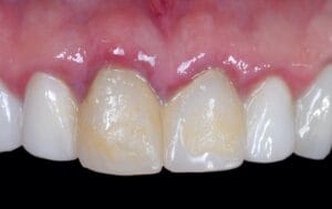
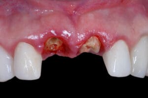
A male, 39-year-old patient presented to the practice with two failed central incisors, which had been the subject of repeatedly failing restorative procedures. The patient sought a permanent solution that would restore function and aesthetics to his central incisors.
Assessment
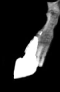
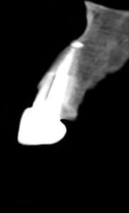
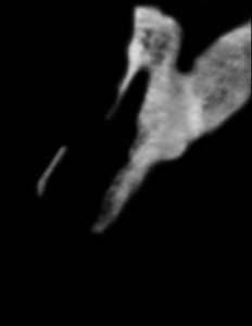
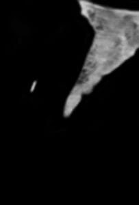
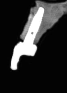
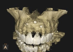
The patient’s medical history demonstrated he was free from any systemic health concerns, and was generally fit and healthy, and a non-smoker. A comprehensive clinical assessment followed, which showed excellent oral hygiene and healthy gingivae. The CT scan taken of the anterior region revealed a significant loss of the buccal plate.
All potential treatment options were discussed with the patient, including his preferred option of implant placement and crowns. Due to the lack of buccal plate and the drawbacks caused by treatment delay until this point, bone augmentation was indicated to ensure primary stability and long-term survival of the implants.
Treatment planning
Following classic treatment protocols, this case would have required delayed implant placement. This could have led to serious aesthetic defects, such as the significant loss of the papillae, post-restorative recession, and scar tissue formation.
However, the author has pioneered a new technique for such clinical situations that overcomes these challenges. VST allows the perfect restoration of the socket in just eight weeks.i
The novel approach supports immediate implant placement with a vestibular incision and cortical bone shield stabilisation, alongside bone augmentation with a particulate bone grafting material. Treatment is complete with a customised healing abutment, enabling the clinicians to provide the entire workflow in a single visit and with a single surgery.
All the benefits, risks and limitations were explained to patient in detail to obtain informed consent. The importance of ongoing oral hygiene for implant longevity was impressed upon the patient at the same time.
Treatment delivery
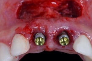
Upon his return to the practice, the patient was numbed with appropriate anaesthesia. The failed restorations were removed to reveal the existing tooth roots, which now had a hopeless prognosis after several failed attempts at restoration.
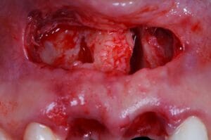
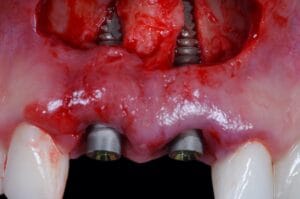
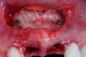
The vestibular access incision was made as per the VST protocol, providing clear visualisation of substantial buccal bone defect. This was confirmed by an intraoperative CBCT scan, ensuring the clinician had comprehensive knowledge of the exact anatomy.
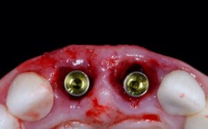
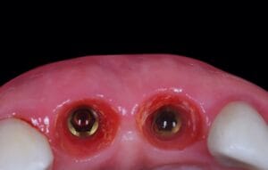
Two 3.8 x 15mm Tapered Pro Conical implants (BioHorizons) were placed. These implants feature a Laser-Lok®/RBT surface, which supports connective tissue attachment and crestal bone volume. They also ensure excellent primary stability, which is crucial in immediate cases. The implants were fully seated, confirming their optimal 3D positioning.
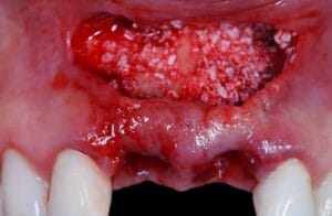
To further promote stability and longevity of treatment, the osseous defect was filled with particulate bone graft, which contained a 50:50 mix of MinerOss® X cancellous particles (BioHorizons) and autogenous cortical bone chips. This combination was chosen to provide optimal regeneration of viable bone.
A cortical membrane was customised and placed on top of the grafting material, stabilised with three membrane tacs to the surrounding bone, using the BioHorizons AutoTac system kit.
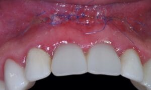
The site was sutured closed tension-free and the patient given standard post-operative care instructions to maintain the health of the site for optimal healing.
Surgical review
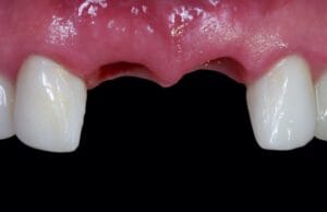
The patient returned to the practice one week post-surgery for review. The site presented a reasonable healing outcome following the immediate non-functional loading protocol.
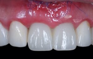
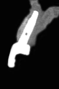
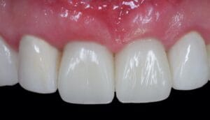
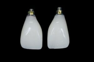
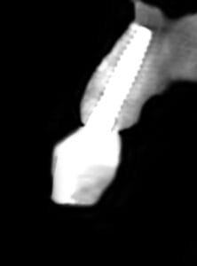
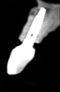
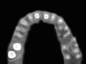
Upon further review post-healing, the buccal contour and papillary heights were effectively maintained, ensuring sufficient soft tissue support and excellent aesthetics. Treatment was concluded with two screw-retained zirconia crowns fitted on two hexed hybrid base abutments from BioHorizons. These provided a final restoration to the previously defective locations, and a superior aesthetic outcome. Two-year follow-up radiographic and CBCT scans demonstrate the volume of regenerated bone two years achievable when using the VST technique.

For product information from BioHorizons, please visit https://theimplanthub.com/
 Author bio: Dr. Abdelsalam Elaskary is the founder of the Vestibular Socket Therapy, as well as the Elaskary & Associates Clinic and Educational Institute. He is also currently a visiting lecturer at University of New York NYU, and the author of three textbooks in the field of dental Implantology and oral reconstruction, published by John Wiley. In addition, Dr Elaskary is currently the President of the Arab Society of Oral Implantology.
[i] Elaskary A, Gaweesh YY, El Tantawi M, Maebed MA. Vestibular Socket Therapy: A Novel Approach for Implant Placement in Defective Fresh Extraction Sockets With or Without Active Socket Infection (One-Arm Cohort Study). Int J Oral Maxillofac Implants. 2021 Jan-Feb;36(1):146-153. doi: 10.11607/jomi.8732. PMID: 33600536.
[ii] ElAskary A, Elfana A, Meabed M, Abd-ElWahab Radi I, Akram M, Fawzy El-Sayed K. Immediate implant placement utilizing vestibular socket therapy versus early implant placement with contour augmentation for rehabilitation of compromised extraction sockets in the esthetic zone: A randomized controlled clinical trial. Clin Implant Dent Relat Res. 2022 Oct;24(5):559-568. doi: 10.1111/cid.13120. Epub 2022 Jul 10. PMID: 35811090.
Other references:
Th Elaskary A, Y Gaweesh Y, Maebed MA, Cho SC, El Tantawi M. A : A Novel Method for Immediate Implant Placement in Defective Fresh Extraction Sites. Int J Oral Maxillofac Implants. 2020;35(4):799-807.
Elaskary A, Meabed M, Abd-ElWahab Radi I. Vestibular socket therapy with immediate implant placement for managing compromised fresh extraction sockets: A prospective single-arm clinical study. Int J Oral Implantol (Berl). 2021 Aug 20;14(3):307-320.
Th Elaskary A, Gaweesh YY, El Tantawi M, Maebed MA. Vestibular Socket Therapy: A Novel Approach for Implant Placement in Defective Fresh Extraction Sockets With or Without Active Socket Infection (One-Arm Cohort Study). Int J Oral Maxillofac Implants. 2021;36(1):146–153.
Elaskary A, Ghallab N : Vestibular socket therapy a novel protocol for immediate imp placenta in compromised fresh extraction sockets : A 3 years floow up case series. Abstracts of EuroPerio10, Copenhagen, Denmark, 15-18 June 2022. J Clin Periodontol. 2022 Jun;49 Suppl 23:4-385
Dhande SH, Elaskary A, Muglikar S, Sheikh S, Hegde R, Kale R, Jabade B, Sayyed S. Treat and Restore Compromised Sockets Immediately with the Novel Vestibular Socket Therapy: A Revolution in Immediate Implant Placement. Glob. J. Med. Res. 2022;21:1-0.
Ghallab N · Elaskary A · Elsabagh H · Toukhy A· Abdelrahman H · El-Kimary G . A novel atraumatic extraction technique using vestibular socket therapy for immediate implant placement: a randomized controlled clinical trial . Oral and Maxillofacial Surgery https://doi.org/10.1007/s10006-022-01089-415 March 2022 / Accepted: 5 June 2022
Elaskary A, Abdelrahman H, Elsabagh H, El-Kimary G : Does Grafting The Jumping Gap In Immediately Placed Anterior Implants Using Vestibular Socket Therapy Influence The Labial Bone Thickness?, Journal of Oral and Maxillofacial Surgery, accepted May 2022,
ElAskaryA , Elfana A, Meabed M, Abd-ElWahab I , Akram M , Fawzy K : Immediate implant placement utilizing vestibular socket therapy versus early implant placement with contour augmentation for rehabilitation of compromised extraction sockets in the esthetic zone: A randomized controlled
clinical trial. Clin Implant Dent Relat Res. 2022;1–11. Accepted: 20 June 2022
Elaskary A, Abdelrahman H, Elfahl B, Elsabagh H, El-Kimary G, Ghallab NA. Immediate Implant Placement in Intact Fresh Extraction Sockets Using Vestibular
Socket Therapy Versus Partial Extraction Therapy in the Esthetic Zone: A Randomized Clinical Trial. Int J Oral Maxillofac Implants. 2023 May, Jun;38(3):468-478.
Hamed MM, El-Tonsy MM, Elaskary A, Abdelaziz GO, Saeed SS, Elfahl BN. Effect of three different grafting materials on immediate implant placement using vestibular socket therapy in class II extraction sockets in the maxillary esthetic zone: a randomized controlled clinical trial. BMC Oral Health. 2023 Sep 1;23(1):623.
Elaskary A, Ghallab N, Thabet A, Shemais N. The bone shielding versus dual-zone concept in treating thin-walled fresh extraction sockets with immediate implant placement: Soft and hard tissue changes. A randomized clinical trial. Clin Implant Dent Relat Res.


