







Author: Nimisha Nariapara – Trade Marketing Manager at Carestream Dental covering the UK, Middle East, Nordics, South Africa, Russia and CIS regions.
Dental implant placement is a complex task, and is only made more difficult when a patient has insufficient bone volume to support a restoration. In many cases, augmentation of the hard tissue is often indicated for treatment predictability and longevity and, depending on the affected site, this may mean the need for sinus augmentation, or a sinus lift.
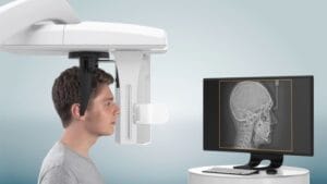
Such a procedure requires clinical skill, experience, knowledge, and a detailed treatment planning process. Maxillary sinus augmentation via the lateral window technique, used for over 40 years, is deemed a safe and predictable surgery.[i] Risk factors such as the presence of septa, thin membrane, gingival biotype, and smoking habits can all be mitigated, resulting in minimal trauma and invasiveness.i
To create a predictable and effective workflow, dental professionals need to engage with high-quality technological systems. Understanding how different solutions optimise the workflow, making any given aspect more efficient or reliable, allows clinicians to choose key investments for their practice that better support their patients.

A clear idea
Long before treatment begins, clinicians need to identify patients suited to sinus augmentation prior to implant placement. After tooth loss, the alveolar ridge is prone to atrophy and pneumatisation of the maxillary sinus.[ii] When a patient loses a tooth in the maxilla and is interested in receiving a dental implant, their clinician will then have to consider the potential for atrophy, and therefore the need for augmentation.
If the clinician is trained, competent and confident to provide this care, then they can begin more detailed assessments; if not, a referral is appropriate,[iii] but the clinician may request further information, such as radiographic images. A presurgical evaluation relies on computed tomography (CT) or cone-beam CT (CBCT) scans, providing a range of information such as membrane thickness, residual bone height, the presence of sinus septa, and the positioning of teeth and other key anatomical features.[iv] These are considered essential before beginning this form of treatment,[v] and clinicians should once again only interpret the results if they have the appropriate training.
With accurate readings and information about the treatment site, the clinician leading the case can proceed with confidence. When investing in an imaging system, it’s important to consider aspects such as accuracy to small scales, which ensures treatment plans are appropriately informed, and wider fields of view that allow for all biological details to be assessed in minimal scans, reducing radiation exposures for patients. Once high-quality scans are secured, clinicians can apply them in many ways, from complete hands-on treatment planning, to artificial intelligence (AI) models, or even CAD/CAM surgical guides.
Digitally-devised assistance
When planning and performing complex dental surgeries, it’s vital that clinicians do not go in blind. Anatomical structures such as nerve canals, nearby teeth, and the skeletal layout of the jaw itself must be managed carefully for a successful outcome. Careful implant planning also allows clinicians to prepare where the restoration will lie, maximising recovery in healthy osseous tissue, and maximising function and aesthetics.
A digitally produced surgical guide has been referenced throughout the available literature as an approach that effectively facilitates minimally invasive surgery. Though it should be known that this is not through removing less tissue, for example by creating a smaller window size or reducing the flap extension, but instead by enhancing the accuracy of the window preparation.[vi] This means that the extent of the sinus membrane elevation needed during surgery could be limited only to the area where the implant will be placed.vi Conventional surgical guides may, however, be fairly large, and demand an excessive reflection of the flap; ensuring this is as small as possible, whilst maximising accuracy is key.[vii]
Clinicians seeking to utilise a guided workflow should seek out systems that maximise accuracy, but also can be implemented seamlessly into the workflows of those creating any physical guides – this could be an in-practice or external dental technician, or even through the same supplier that provides digital planning software.
Optimising results
In the future, clinicians will expect to see AI play a key role in sinus augmentation treatment, and implant planning as a whole. This will rely on accurate scans of the maxillofacial structures in order to devise a precise, effective plan.
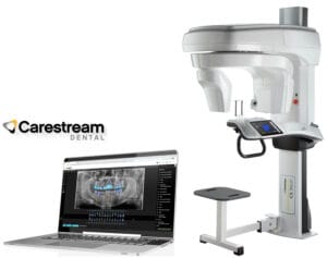
The CS 9600 CBCT Scanner from Carestream Dental is a leading solution for clinicians looking to take on complex cases with confidence. The versatile 5-in-1 scanner delivers smart innovations and precise outcomes, with up to 14 fields of view available for maximum insight into all affected anatomy. Powered by CS 3D Imaging Software now including premium features such as AI automated implant planning to increase efficiency and accuracy, your workflow can be transformed for improved patient outcomes.
A variety of technological solutions can aid the delivery of sinus augmentation and subsequent implant placement. Effective planning can only begin when clinicians have a clear idea of a patient’s condition, making high-quality dental imaging an important place to start.

For more information on Carestream Dental visit www.carestreamdental.co.uk
For the latest news and updates, follow us on Facebook and Instagram @carestreamdental.uk
[i] Valentini, P., & Artzi, Z. (2023). Sinus augmentation procedure via the lateral window technique—Reducing invasiveness and preventing complications: A narrative review. Periodontology 2000, 91(1), 167-181.
[ii] Lyu, M., Xu, D., Zhang, X., & Yuan, Q. (2023). Maxillary sinus floor augmentation: a review of current evidence on anatomical factors and a decision tree. International journal of oral science, 15(1), 41.
[iii] General Dental Council, (2019). Standards for the dental team. (Online) Available at: https://www.gdc-uk.org/standards-guidance/standards-and-guidance/standards-for-the-dental-team [Accessed September 2025]
[iv] Alshamrani, A. M., Mubarki, M., Alsager, A. S., Alsharif, H. K., AlHumaidan, S. A., Al-Omar, A., … & ALHumaidan, S. A. (2023). Maxillary sinus lift procedures: an overview of current techniques, presurgical evaluation, and complications. Cureus, 15(11).
[v] Misăiloaie, A., Tărăboanță, I., Budacu, C. C., & Sava, A. (2024). Preoperative Cone Beam Computed Topography Assessment of Maxillary Sinus Variations in Dental Implant Patients. Diagnostics, 14(17), 1929.
[vi] Sirinirund, B., Rodriguez Betancourt, A. B., Scaini, R., Wu, Y. C., & Chan, H. L. (2025). Minimally Invasive Sinus Augmentation: A Systematic Review. Clinical implant dentistry and related research, 27(1), e13403.
[vii] Cho, S. W., Yang, B. E., Cheon, K. J., Jang, W. S., Kim, J. W., & Byun, S. H. (2020). A simple and safe approach for maxillary sinus augmentation with the advanced surgical guide. International journal of environmental research and public health, 17(11), 3785.


