





Dr Balaji presents a case in which complex soft tissue augmentation was undertaken to address gingival recession at the UL3, UR3, and UR1. In this case, the key aim was to treat the soft tissue to reduce the risk of tooth loss and produce an aesthetic outcome.
Patient presentation and assessment
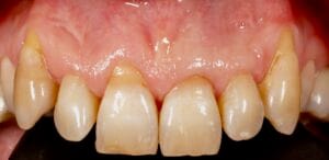
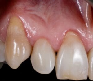
A 58-year-old man presented to the practice for treatment. On assessment, the patient had Miller classification class II recession at the UL3 site, in addition to class II recession at the UR3 and UR1. The prospect of soft tissue augmentation was explained to the patient, and the benefits it offers in terms of reducing the risk of tooth loss.
Treatment planning
Radiographs were taken of the affected areas which showed that, although there was gingival recession, there was no bone loss. It was recommended to complete treatment in two stages, beginning with the UL3 in the first instance and completing treatment at the UR3 and UR1 at a later date.
UL3
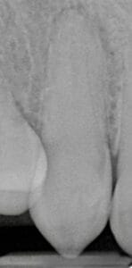
In order to address the gingival recession at the UL3, it was important to discuss the potential treatment options, including doing nothing, which was not recommended in this case.
Soft tissue augmentation was deemed to be the most appropriate treatment pathway in this case. The use of a flap to cover the graft – known as the bilaminar technique – improves predictability of root coverage because it provides the graft with an additional blood supply. Additionally, it offers an aesthetic result by hiding the white scar colour of the graft and masking the irregular outline of the mucogingival junction which is common after a grafting procedure. Research suggests that the bilaminar technique is an effective procedure in the treatment of gingival recession, particularly in patients with aesthetic demands, as it results in complete soft tissue root coverage in the majority of cases.[i]
Additionally, the design of the mucogingival flap is significant, the trapezoidal flap technique consists of two horizontal and two slightly divergent vertical incisions extending to the alveolar mucosa.[ii] This technique is beneficial as the design is such that the base of the flap is wider, making the blood supply more reliable.[iii]
UR3 and UR1
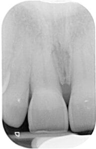
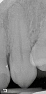
Soft tissue grafting was also recommended for the treatment of gingival recession at the UR3 and UR1. In order to ensure the best results, the patient consented to the placement of connective tissue grafts and soft tissue augmentation to both treat the recession and restore gingival thickness to this area of the mouth.
Treatment provision
UL3
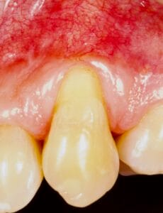
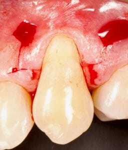
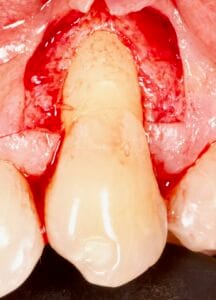
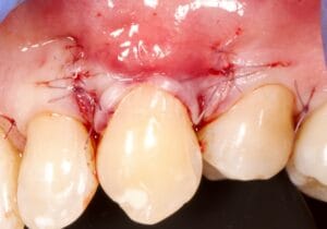
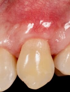
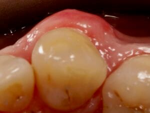
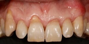
According to the treatment plan, the mucogingival flap was designed and raised, following the trapezoidal flap and ressective technique. The recession at the site was measured and the flap added 1 mm. Once the root was exposed, it was cleaned by root planing, using EDTA and amelogenin.
A connective tissue graft (CTG) was harvested from the palate, and sutured to the canine site. The flap was then advanced to cover the connective tissue graft, following the bilaminar technique. Good coverage was achieved by carrying out this approach, as is reflected in the clinical images. The patient was provided with appropriate post-operative instructions to encourage optimal healing.
UR3 and UR1
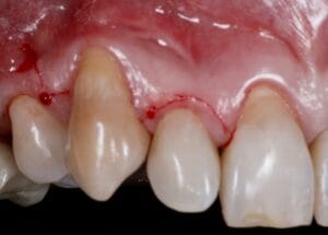
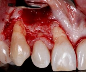
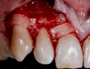
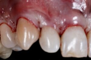
To treat the gingival recession at the UR3 and UR1, a split thickness flap designed for multiple teeth was raised. Gingival planing was then undertaken using EDTA and amelogenin.
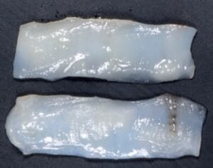
Two CTGs were harvested from the palate and placed onto the root surfaces of the UR3 and UR1 to ensure optimal gingival height is restored. Once stabilised, the flap was advanced tension-free to cover the roots. As before, the patient was given post-operative oral health instructions to facilitate smooth healing.
Reflection
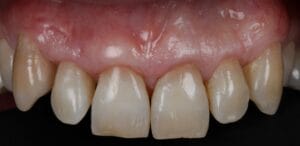
An excellent outcome was achieved in this case, with good soft tissue coverage at the UL3 ensuring an aesthetic result. The main challenge presented was that the tooth was placed quite buccally, which made avoiding perforation difficult during surgery.
Dr Balaji provides industry-leading training courses on both hard and soft tissue management around dental implants with the ASHA Club.
For more information about how you could elevate your skills with the support of experts, please visit www.ashaclub.co.uk or call 07974 304269
 Dr Selvaraj Balaji: BDS, MFDS RCPS(Gla), MFD SRCS(Ed), LDS RCS(Eng)
Since he obtained the BDS Degree, Dr Balaji has worked in Maxillo-facial units in the UK for several years and gained substantial experience in surgical dentistry. He is the principal dentist of The Gallery Dental Group which is made up of Meadow Walk Dental Practice and The Gallery Dental & Implant Centre. Dr Balaji is also the founder of the Academy of Soft and Hard Tissue Augmentation (ASHA) and runs courses, lectures and study clubs in the UK and around Europe for aspiring implantologists.
[i] Zucchelli G, Amore C, Sforza NM, Montebugnoli L, De Sanctis M. Bilaminar techniques for the treatment of recession-type defects. A comparative clinical study. J Clin Periodontol. 2003 Oct;30(10):862-70. doi: 10.1034/j.1600-051x.2003.00397.x. PMID: 14710766.
[ii] Zucchelli, G., Stefanini, M., Ganz, S., Mazzotti, C., Mounssif, I. and Marzadori, M., 2016. Coronally Advanced Flap with Different Designs in the Treatment of Gingival Recession: A Comparative Controlled Randomized Clinical Trial. International Journal of Periodontics & Restorative Dentistry, 36(3).
[iii] Kitcat, M, and S L Benyon. “Trapezoid advancement flap to reconstruct small to medium sized tissue defects-A series of clinical applications.” JPRAS open vol. 33 32-36. 12 May. 2022, doi:10.1016/j.jpra.2022.05.001


