






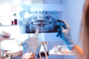
Your patient is somewhat dentophobic, and they’ve been working through their anxieties under your care. They required a restoration to a tooth after developing caries, but have since made efforts to improve their diet and oral hygiene routine, and even managed to remain calm during their appointment where you placed their first (and hopefully last) composite restoration. Delighted with their ability to face their fears, they have diligently looked after their restoration and strived to make more regular dental appointments.
Upon their return to the practice to monitor the health of the new restoration, you take a radiograph. On first appearance your patient has developed secondary caries. You know that this might mean replacement of the restoration completely, and understand how this might impact your patient’s confidence and progress through dentophobia, potentially impacting the progression made in oral health improvements – but can you be certain of what your radiograph is telling you?
Secondary caries diagnosis is sometimes difficult to differentiate from restorative materials or other defects. The literature[i] notes that a highly sensitive approach to detection could identify nearly all lesions, but increase the risk of false-positive diagnoses. On the other hand, a highly specific approach would avoid overtreatment, but could miss many incidences of secondary caries, leaving the restoration to suffer as a result. Understanding why it can be difficult to identify secondary caries is key to providing patients with the highest quality of care.
Crown and composite confusion
Around 50-60% of defective restorations are replaced due to secondary caries, but this could be a clear case of overtreatment, since the rate of restoration failure due to secondary caries is as low as 2-3% in controlled clinical trials.[ii] Visual misdiagnoses may lead to clinicians confusing secondary caries lesions with microleakage or residual caries, and radiographic assessments can lead to professionals mischaracterising restorative material radiopacity, the presence of bond layer, or residual caries.ii
The radiopacity of restorative materials is especially important to focus on. This feature enables clinicians to more easily distinguish restoration from dental defects, minimising the risk of overtreatment due to misdiagnosis of an issue such as secondary caries. Many restorations and dental cements are radiopaque naturally – especially amalgam or metal alloys – and composite materials often use specific fillers to increase their visibility in X-ray images.
This isn’t always the case with dental adhesives. When a clinician uses a solution with insufficient radiopacity, and then observes a radiograph, they can face interpretation struggles; radiolucency below a composite restoration could be secondary caries, a lack of marginal adaption, or simply a thick layer of adhesive material. To prevent overtreatment, a clinician needs to monitor the patients’ health over time, taking action when clinical and radiographic changes are observed in ensuing visits. How the clinician places the initial restoration can also make a difference – demanding clinical skill and the correct restorative solutions to achieve an optimal result.
In the clinician’s hands
Secondary caries can appear below a restoration for a number of reasons. Sometimes called recurrent caries, development is a multifactorial process which takes on the key pathogenesis for ‘conventional’ caries, but is modified by a restoration or sealant margin.[iii] Since the problem is partially linked to the quality of the restoration, the clinician can be a main factor behind secondary caries development – and in turn, a key contributor to a restoration’s success.i
The dental professional must adequately remove residual caries, and apply restorative materials at an adequate depth and in a sufficient quantity. Gaps beneath restorations may be a result of poor initial placement of a restoration, and can cause subsequent issues potentially requiring retreatment.i
Clinicians also, of course, have control over the materials used. When choosing those for a restoration, it’s important to not only identify the best possible solutions for an immediate success, nor one that will last long into the future, but recognise that upon reassessment, the radiopacity of the materials chosen can affect treatment choices.
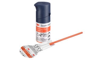 An adhesive solution like the 3M™ Scotchbond™ Universal Plus Adhesive from Solventum is ideal for a range of indications. Offering control and predictability, the material provides gold standard adhesion to all dental substrates, and bonds and seals caries affected dentine to support minimally invasive dentistry. Importantly, it is also the first universal adhesive with dentine-like radiopacity – this reduces the risk of misdiagnosis and overtreatment down the line, supporting your patients and the clinicians that care for them.
With the recognition that dental materials, and therefore the choices of the clinician, can affect the risk of overtreatment, dental professionals should endeavour to have reliable and effective solutions on-hand. Increased radiopacity can help clinicians when making assessments in the future, and in turn give patients an improved chance at high-quality care.

To learn more about Solventum, please visit https://www.solventum.com/en-gb/home/oral-care/
For more updates on trends, information and events follow us on Instagram at @solventumdentalUK and @solventumorthodonticsemea
©Solventum 2024. Solventum and the S logo are trademarks of Solventum and its affiliates. 3M and Scotchbond are trademarks of 3M company.
About Solventum
Solventum, enabling better, smarter, safer healthcare to improve lives. As a new company with a long legacy of creating breakthrough solutions for our customers’ toughest challenges, we pioneer game-changing innovations at the intersection of health, material and data science that change patients’ lives for the better while enabling healthcare professionals to perform at their best. Because people, and their wellbeing, are at the heart of every scientific advancement we pursue. We partner closely with the brightest minds in healthcare to ensure that every solution we create melds the latest technology with compassion and empathy. Because at Solventum, we never stop solving for you.
[i] Askar, H., Krois, J., Göstemeyer, G., Bottenberg, P., Zero, D., Banerjee, A., & Schwendicke, F. (2020). Secondary caries: what is it, and how it can be controlled, detected, and managed?. Clinical oral investigations, 24(5), 1869-1876.
[ii] Signori, C., Gimenez, T., Mendes, F. M., Huysmans, M. C. D., Opdam, N. J., & Cenci, M. S. (2018). Clinical relevance of studies on the visual and radiographic methods for detecting secondary caries lesions–A systematic review. Journal of Dentistry, 75, 22-33.
[iii] Askar, H., Krois, J., Göstemeyer, G., Bottenberg, P., Zero, D., Banerjee, A., & Schwendicke, F. (2020). Secondary caries: what is it, and how it can be controlled, detected, and managed?. Clinical oral investigations, 24(5), 1869-1876.


