






Dr Paul Swanson presents a case that required the replacement of both missing upper canines, giving this young patient her smile and her confidence.
Introduction
Although it is uncommon for dental professionals to consider implant treatment for young patients, there are situations where it is appropriate. Some patients with congenitally missing teeth may be ideal candidates.
Approximately 3-5% of the UK population experience hypodontia,[i] with treatment usually involving the orthodontic closure of spaces or orthodontic widening of gaps to allow replacement of the missing tooth. One option for the replacement of a congenitally absent tooth is dental implant placement. The following case is an example of the latter, with a young patient looking to complete two years of alignment with replacement of her missing canines.
Patient presentation
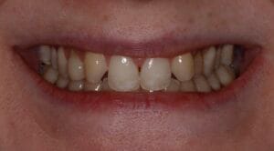
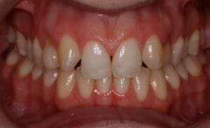
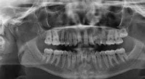
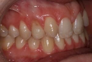
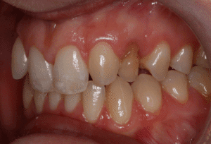
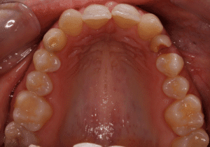
A young female patient presented to the practice with congenitally absent upper lateral incisors. She was 25 at the time of presentation. Her upper canines were positioned in the sites of the lateral incisors with small spaces between these and the upper pre-molars causing misalignment. Radiographs showed significant bone resorption at the canine sites, as would be expected. As a result, the patient was warned about the potential need for complex bone augmentation prior to implant placement. It would be necessary to reassess the bone volume at the end of orthodontic treatment.
The patient then proceeded to undergo approximately two years of orthodontic treatment. This had been designed to align both arches, while creating good occlusion and adequate space for implant placement in the canine sites.

Assessment
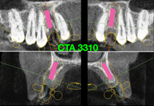
Throughout orthodontic treatment, good communication was maintained between the orthodontist and implant dentist. This was to ensure that the correct space was created both coronally and apically for implant placement. A comprehensive assessment was performed to determine the most effective approach to implant treatment post-orthodontics. A CBCT scan was taken to evaluate the existing bone quality, as was previously planned. This established that augmentation would be possible alongside implant placement, but accuracy of implant positioning would be crucial.
Treatment plan
The clinical decision was made not to ‘distalise’ the canines but instead to move them laterally and focus on implant placement in the canine spaces.
The radiographic images were used to plan implant placement using SMOP design software. It was determined that narrow conical implants of 3.3mm in diameter could be used in order to facilitate implant placement with simultaneous bone augmentation. A guided approach would also be utilised to increase accuracy of implant placement and enhance the chance of success.
A digital workflow was implemented to identify the ideal implant positions and design the surgical guide. Intraoral scans and all other appropriate diagnostic images were shared with the laboratory for the fabrication of the guide.
All the benefits and risks of the proposed treatment were once again discussed with the patient to ensure she understood exactly what to expect and obtain informed consent.
Treatment
Surgical treatment commenced with the raising of a flap to provide visualisation of the two canine sites.
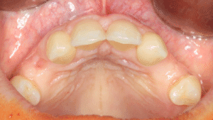
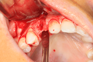
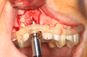
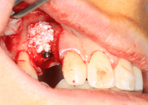
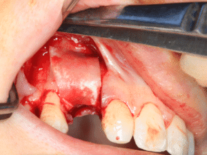
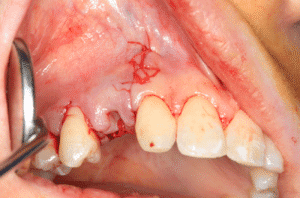
Two 3.3mm diameter Tapered Pro Conical implants (BioHorizons Camlog Group) were then placed using a fully guided approach to ensure precision. These narrow implants are very useful in cases such as this because they allow placement in sites with limited mesiodistal space, but demonstrate good strength characteristics in comparison to other larger implants on the market. The implants were placed entirely into bone, with contour augmentation carried out to increase the buccal bone thickness and improve the contour of the labial surface. MinerOss® X (BioHorizons Camlog Group) bovine bone mineral was used – the benefit of this solution is its ability to not resorb and therefore maintain volume in the long-term. It was held in place with a Mem-Lok® membrane, which was secured with bone tacks to increase the stability and predictability of the graft.
A 2mm height abutment was placed to support the bone augmentation and aid the second-stage surgery. The site was sutured closed tension-free and the patient given post-operative hygiene and care instructions.
The patient returned to the practice three months later. The implants were exposed during the second-stage surgery to reveal healthy soft tissues. The lab-made provisional crowns were placed on the UL3 and UR3. The final restorations were cement-retained crowns connected to customised DEDICAM abutments (BioHorizons Camlog Group).
Outcome and discussion
The patient was delighted with the outcome achieved in this case, happy to finally have the canines replaced and be able to show off a complete smile. She has since returned for a new orthodontic retainer to maintain the tooth positions and protect her new crowns.
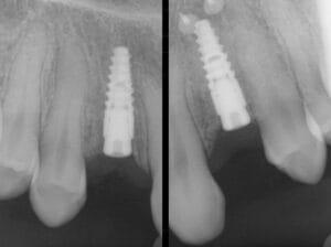
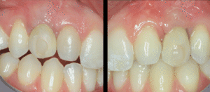
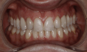
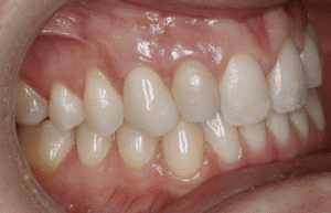
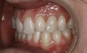
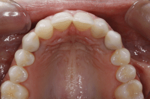
Although the general preference is for screw-retained implant crowns, cement-retained options were chosen for this case. The patient had limited available bone, and placing implants within the limited bone meant the fixtures were angled, with the apex of the fixtures into the palatal bone. This prevented the need for a substantial bone graft prior to implant placement, potentially making the treatment journey more efficient and comfortable for the patient.
Success in this case meant ensuring adequate distance between the implants and the neighbouring natural teeth. It was also crucial to make sure that the implants could be placed into bone, and so selection of the augmentation materials was key to optimising the buccal bone thickness. Another essential factor was the treatment planning for the 3D positioning of the implant. As the implants were angled into the palatal bone, it was important to achieve the correct depth of placement (deeper in this case) as this had a significant impact on the aesthetics that could be achieved with cement-retained crowns by giving ‘space’ for the customised abutments.
For product information from BioHorizons Camlog Group, please visit https://theimplanthub.com/
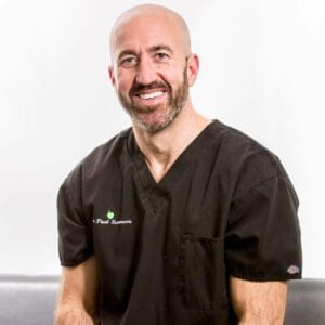 Author bio: Dr Paul Swanson has been the principal of Rose Lane Dental Practice in Liverpool. Following his qualification from the University of Liverpool in 2000, Paul continued his studies to obtain a Diploma in General Practice from the Royal College of Surgeons in 2003. In 2008 he achieved the Diploma in Implant Dentistry and Advanced Certificate in Bone Grafting from the Royal College of surgeons of England. In 2010 he received an MSC in Implant Dentistry from Queen Mary University of London.
Paul accepts referrals from other practices across the city and also further afield for implant dentistry. He has previously been employed by Liverpool University Dental Hospital where he helped train and mentor newly graduated dentists and was a tutor with the Royal College of Surgeons Implant Diploma.
[i] NHS. Missing teeth (hypodontia). https://www.nuh.nhs.uk/orthodontics-missing-teeth-hypodontia/ [Accessed March 2025]


