






Despite endodontic treatment being one of the more technically demanding procedures in dentistry, the success rates are proven to be as high as 85.2-92.6% for primary root canal treatment[i][ii] and 77.2-83% for root canal retreatment.[iii]
Successful endodontic treatment can maintain long-term oral health by preserving natural teeth, alleviating patient pains and complaints. Despite this, endodontic procedures still face many different challenges, meaning failures remain a significant hurdle to overcome. This can lead to retreatments, infections, and even tooth loss that must then be rectified with dental implants and such.[iv]
Although materials, technology, and techniques surrounding endodontics have progressed, the achievement of consistent, predictable, and long-lasting root canal therapy success remains difficult. To minimise the risk of failure, clinicians must implement specific protocols and optimise the advantage of modern tools to support each stage of the treatment procedure.
Endodontics failure factors
Many dentists feel apprehensive about performing endodontic treatment due to the technical difficulty, intense training, and greater risk of complications that come with it. Even the smallest errors can cause patient dissatisfaction, which poses the threat of legal action. This fear of litigation can often lead to complex cases being referred to specialists.[v]
When endodontic treatment fails, it can be for a plethora of reasons. One step that commonly jeopardises success is the inability to properly locate, clean, and fill all the canals in the root.[vi] The incomplete debridement of the root canal system leaves residual bacteria or necrotic tissue behind and cause excruciating pain, or even lead to ongoing periapical disease.[vii][viii] Inadequate obturation also leads to reinfection, meaning compromised treatment and prolonged issues.[ix]
It is particularly common to miss a canal during endodontic treatment when treating molar teeth. The issue stems from the frequent misunderstanding that there is simply one root and one canal, but molars often have more canals than roots.[x] One rife but underestimated factor is the incorrect determination of working length. Without recognition of the precise working length, clinicians risk failing to clean the canal system entirely, which increases the likelihood of treatment failure considerably.[xi]
The importance of accurate working length determination
Establishing the correct working length is imperative for the success of root canal therapy. It essentially sets the parameters for cleaning and obturation, and miscalculations in such can compromise the entire treatment.
Over-instrumentation often causes irrigants or other substances to extrude beyond the apex, causing periapical damage and/or constant inflammation that leads to extreme patient discomfort.[xii] Opposingly, under-instrumentation leads to loss of working length, and the lingering effects of residual bacteria can cause enduring failure and long-lasting, painful infection.[xiii]
There are multiple methods that clinicians use in establishing the working length of root canals which include radiographic, tactile, and electronic. Arguably the most popular and traditional approach leans heavily upon periapical radiographs.[xiv] However, these are not especially reliable due to their inability to provide anything more than a two-dimensional view of a three-dimensional structure. Hence, they don’t have the capability to capture canal curvature or apical anatomy. Depending solely on radiographs can increase the risk of misjudging canal length due to inaccurate readings, particularly in complex cases.[xv]
Tools differentiate success and failure
The endodontic workflow has advanced due to substantial technological developments which have improved both precision and efficiency. Canal detection has improved with the introduction of dental microscopes and loupes, while ultrasonic activation and other methods of assisting irrigation help to enhance the cleaning of the root canal.[xvi][xvii]
Additionally, clinicians’ incorporation of electronic apex locators into their routines is now a common part of endodontic practice, revolutionising assessments to ensure working length accuracy. The implementation of these modern technologies plays a vital role in promoting long-lasting success by greatly reducing the risk of endodontic failure.
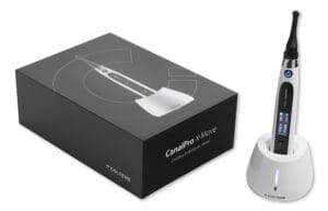 One tool that allows clinicians to accurately establish working length is the CanalPro® X-Move from COLTENE. With an integrated apex locator, it delivers true, dependable measurements that remove the reliance on radiography. Its innovative technology produces consistent readings even in the most complex of cases.
The CanalPro® Apex Locator is intuitive and user-friendly, augmenting clinicians’ confidence in determining accurate working length and preparing root canals. By improving precision at this vital step in the process, clinicians can reduce the risk of over- or under-instrumentation, improving results holistically.
Improving your chances
Avoiding endodontic failure is dependent upon the meticulousness of each step in the treatment process, as well as support from dependable technologies. Clinicians who invest in accurate, proven tools, rather than relying on those that cannot guarantee consistent triumph, will inevitably meet success.

For more information, visit https://colteneuk.com/CanalPro_X-Move
email info.uk@coltene.com or call 0800 254 5115.
 Author: Nicolas Coomber – COLTENE National Account & Marketing Manager
[i] Ng, Y. L., Mann, V., Rahbaran, S., Lewsey, J., & Gulabivala, K. (2007). Outcome of primary root canal treatment: systematic review of the literature – part 1. Effects of study characteristics on probability of success. International endodontic journal, 40(12), 921–939. https://doi.org/10.1111/j.1365-2591.2007.01322.x
[ii] Burns L E, Kim J, Wu Y, Alzwaideh R, McGowan R, Sigurdsson A. Outcomes of primary root canal therapy: an updated systematic review of longitudinal clinical studies published between 2003 and 2020. Int Endod J 2022
[iii] Ng Y-L, Mann V, Gulabivala K. Outcome of secondary root canal treatment: a systematic review of the literature. Int Endod J 2008
[iv] Daokar, Sadashiv. (2013). Endodontic Failures-A Review. IOSR Journal of Dental and Medical Sciences. 4. 5-10. 10.9790/0853-0450510.
[v] Morgan, A., Youngson, C. & McLean, W. Medico-legal considerations in endodontics. Br Dent J 238, 573–579 (2025). https://doi.org/10.1038/s41415-025-8333-z
[vi] Abdulwahab, M.A. et al. (2021) ‘Persistence of bacteria and its role in Endodontic treatment failure’, International Journal Of Community Medicine And Public Health, 9(1), p. 432. doi:10.18203/2394-6040.ijcmph20214834.
[vii] Rouhani, A., Aboutorabzadeh, S. M., Reyhani, M., Kheirabadi, N., Mortazavi, S., & Navabi, S. (2023). Prevalence of missed canals in endodontically treated teeth: A cone-beam computed tomography study. Journal of clinical and experimental dentistry, 15(8), e605–e611. https://doi.org/10.4317/jced.60282
[viii]Tabassum, S., & Khan, F. R. (2016). Failure of endodontic treatment: The usual suspects. European journal of dentistry, 10(1), 144–147. https://doi.org/10.4103/1305-7456.175682
[ix] Tabassum, S., & Khan, F. R. (2016). Failure of endodontic treatment: The usual suspects. European journal of dentistry, 10(1), 144–147. https://doi.org/10.4103/1305-7456.175682
[x] Tabassum, S. and Khan, F.R. (2016a) ‘Failure of endodontic treatment: The usual suspects’, European Journal of Dentistry, 10(01), pp. 144–147. doi:10.4103/1305-7456.175682.
[xi] Sethi, K., Shefally, S., & Raji, J. (2021). Working length determination: A review. International Journal of Health Sciences, 5(S1), 45–55. https://doi.org/10.53730/ijhs.v5nS1.5309
[xii] Shacham, M., Levin, A., Shemesh, A. et al. Accuracy and stability of electronic apex locator length measurements in root canals with wide apical foramen: an ex vivo study. BDJ Open 6, 22 (2020). https://doi.org/10.1038/s41405-020-00052-3
[xiii] Chandler, N., Alothmani, O. and Friedlander, L. (2013) ‘The anatomy of the root apex: A review and clinical considerations in Endodontics’, Saudi Endodontic Journal, 3(1), p. 1. doi:10.4103/1658-5984.116273.
[xiv] Abdulwahab, M.A. et al. (2021) ‘Persistence of bacteria and its role in Endodontic treatment failure’, International Journal Of Community Medicine And Public Health, 9(1), p. 432. doi:10.18203/2394-6040.ijcmph20214834.
[xv] Sarsam, W., Davies, J. & Al-Salehi, S. The role of imaging in endodontics. Br Dent J 238, 448–457 (2025). https://doi.org/10.1038/s41415-025-8511-z
[xvi] Low, J. F., Dom, T. N. M., & Baharin, S. A. (2018). Magnification in endodontics: A review of its application and acceptance among dental practitioners. European journal of dentistry, 12(4), 610–616. https://doi.org/10.4103/ejd.ejd_248_18
[xvii] Mozo, S., Llena, C., & Forner, L. (2012). Review of ultrasonic irrigation in endodontics: increasing action of irrigating solutions. Medicina oral, patologia oral y cirugia bucal, 17(3), e512–e516. https://doi.org/10.4317/medoral.17621


