Planmeca decreases patient doses for 3D images of the head – algorithms provide more detailed imaging and diagnostics
NewsPosted by: Dental Design 3rd July 2020
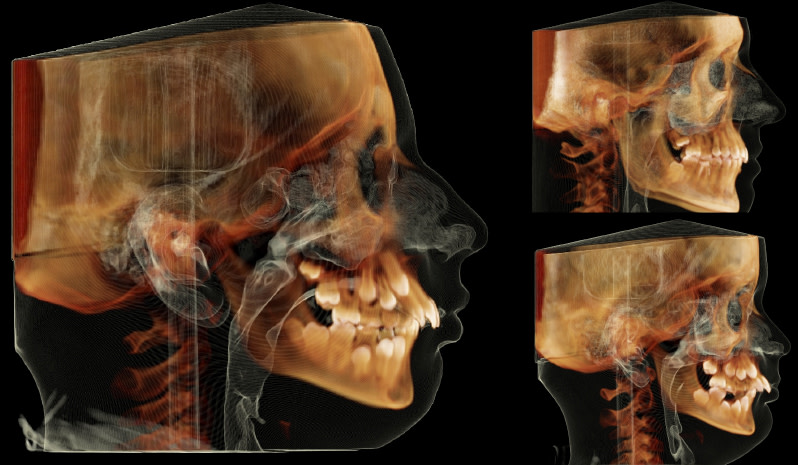
Planmeca has developed smart technologies that help decrease the amount of radiation required to produce medical images without compromising the diagnostic quality of the images. The algorithms developed for 3D imaging help produce more detailed images and diagnostics. Low-dose imaging is beneficial for any patient, but it is particularly significant for children, as a child’s body has comparably more radiation-sensitive red bone marrow than the body of an adult.
When new scientific studies on the subject were published around a decade ago, Planmeca began to develop its own dose measurement solutions. Planmeca believed that these solutions could help decrease ionising radiation doses in medical imaging.
Juha Koivisto, Planmeca’s Chief Physicist and a long-time employee, decided to write his doctoral dissertation on the subject at the University of Helsinki – We noticed during our research that we could decrease the radiation dose without affecting the diagnostic quality of our images. When we compared our low-dose imaging solution to traditional methods used in the field, we discovered that the radiation dose could be reduced by 77%, says Koivisto.
Using a dosimeter to observe how radiation is formed
Planmeca Ultra Low Dose is a smart, algorithm-based low-dose imaging protocol that was launched in 2014. Before the company could develop the new product, it first needed to create a real-time dosimeter. The device allows the company’s researchers to observe how a radiation dose is formed.
– The dosimeter includes a patient model called a phantom, which has sensors that measure radiation doses. When a phantom is exposed to a radiation beam produced by a CBCT unit or another X-ray device, these sensors measure residual dose levels in tissues in order to derive effective doses. This allows us to gain an understanding of the organs the beam reaches and the makeup of the effective dose in the head region. Different organs have different weighting factors, notes Juha Koivisto.
For example, bone marrow, which is renewed at the fastest rate, has the largest weighting factor. If it is exposed to radiation, the effective dose is larger than a dose that is aimed at skin or the brain, Koivisto continues.
The dosimeter was used to review every imaging protocol used by Planmeca.
Diagnostic requirements determine the area, precision and radiation dose of an image
Planmeca is developing its clinical 3D imaging applications for implant and orthodontic treatments, respiratory tract and sinus examinations, and maxillofacial surgeries.
With Planmeca’s devices, dentists and radiologists can now base the right imaging protocols and radiation doses on each patient’s diagnostic needs. Professionals in the field usually have a good understanding of the radiation doses required to achieve images of a certain quality. The Finnish Radiation Act mandates that a patient should not be subjected to more than the minimum amount of radiation necessary for the diagnosis in question – If, for example, one wants to diagnose a patient’s frontal or maxillary sinuses, a smaller radiation dose is usually enough to obtain a sufficiently detailed image, notes Koivisto.
Implant or root area treatments require more enhanced and detailed 3D images, and the imaging of these smaller regions often requires larger radiation doses. However, the jaw area images used as part of orthodontic treatments usually require images of a lower resolution and a smaller radiation dose.
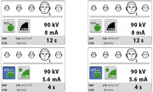
Planmeca’s devices can capture images of a 10 X 10 cm area with a radiation dose as low as 12 microsieverts. Physicians can lower the dose by, for example, limiting the size of the volume or by decreasing the imaging values used, i.e. by changing the quality of the image. In practice, the low dose imaging process has been made to be as quick and easy as possible – all you need to do is press a green button.
The goal is to decrease cumulative radiation exposure in the populace
Juha Koivisto explains that the statistical risk of radiation is very small for an individual, but this figure can still be significant at the wider population level – We want to further decrease the cumulative risk that radiation poses to the populace and we can achieve this by decreasing the radiation doses of our devices to a level that is as low as possible, he notes.
Planmeca’s devices can capture a medical 3D image with a dose as low as 12 microsieverts, which is the equivalent of four days of background radiation in Finland – According to the Radiation and Nuclear Safety Authority, we are exposed to around 0.15 microsieverts of radiation per hour. For example, on a transatlantic flight you are exposed to around 80 microsieverts of radiation, which is significantly higher than the lowest radiation dose produced by CBCT imaging, Koivisto describes.
Planmeca’s objective is to decrease the radiation doses of its devices even further, to the lowest levels possible, in accordance with the ethical standard of “as low as diagnostically acceptable” (ALADA). This principle is part of Planmeca’s ethical imaging philosophym– Even though an individual patient is not subjected to any great risks when an image is captured of their head region, on a wider scale this can become a national health issue, which is why it is advisable to further decrease radiation doses, Juha Koivisto says.
New principles inspire debate
The European Commission drafts evidence-based guidelines on which imaging protocols are to be used for different cases, and it also publishes the average radiation dose values used in medical imaging.
Antti Airisto, an Area Export Manager at Planmeca, explains that these policies inspire a great deal of debate among professionals in the field. Today, medical images are acquired using both 2D and 3D imaging devices, which also affects the average values.
There has also been discussion, for example, surrounding the use of 3D imaging for wisdom tooth removal cases. Among other things, 3D images can help reveal cases where the root of a patient’s tooth is tangled around a nerve canal.