Preserving natural dental tissue – Justin Smith
Featured Products Promotional FeaturesPosted by: Dental Design 17th June 2019

Preventive dentistry focuses on the procedures and practices that help to inhibit the start or arrest the progression of tooth decay and other oral diseases. For dental professionals, the main aims are to prevent damage or disease by identifying problems at the early stages and to preserve or restore natural tissues without invasive intervention. Nevertheless, when one looks at the scale of the problem, dental decay remains the world’s most widespread disease[1]and poses a significant health issue in the UK. The statistics on childhood tooth decay for example, are particularly alarming, accounting for over 26,000 hospital admissions in 5 to 9 year olds during2017-2018.[2],[3]
The prevention of oral disease encompasses the procedures delivered in practice as well as the provision of education and instruction to help patients to maintain healthy teeth and gums. It is dependant upon the oral hygiene routine performed by patients at home along with their individual behavioural and lifestyle choices. Nonetheless, as our scientific knowledge continues to develop and as technology evolves, advancements are constantly being discovered to help improve the oral health of patients and to assist practitioners in achieving preventative, minimally invasive dentistry objectives.
As dental professionals are aware, once lost, tooth enamel cannot regenerate and discovering a way to successfully recreate a substance that looks, feels and behaves like tooth enamel is a significant requirement in the dental sector. However, it appears that there is light at the end of the tunnel provided by the Queen Mary’s School of Engineering and Materials Science. Hereresearchers and scientists have found a new way of developing synthetic mineralising materials that have a mechanism that triggers and guides the growth of apatite nanocrystals in a similar way to the highly organised structure of natural dental enamel.[4]
In addition, and due to the combined efforts of a team of both British and American researchers, another synthetic biomaterial has also been produced. When it is placed in direct contact with pulp tissue, and exposed to a low-dose laser treatment, scientists have been able to stimulate native stem cells and fill carious lesions with a new, natural bony tissue.[5]Although largely at the experimental stage, the results are extremely promising and it has been demonstrated that stem cells from the back molars or wisdom teethcan beinjected into adecaying tooth to growreplacement tissue and blood vessels.[6]
Also coaxing tooth enamel to regenerate on its own, scientists from the University of California have engineered a string of amino acids that contain only the parts required for enamel crystal creation. This shorter peptide is able to grow hard, synthetic enamel and it is hoped that it can be incorporated into a gel and applied to eroded teeth or those affected by early cavities.[7]
These remarkable breakthroughs open up a wealth of opportunities for dentistry and offer huge potential for addressing conditions such as dentin hypersensitivity, tooth decay and dental erosion.[8]However it may be some time before this type of regenerative technology can be brought to market. Consequently, identifying problems as early as possible remains vital in order to help patients to restore their teeth to health with preventive steps and conservative intervention.
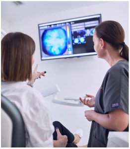
Meeting the demand for minimally invasive dentistry, the CALCIVIS®team have developed a revolutionary new imaging system that successfully identifies and visualises active demineralisation, which is associated with caries and dental erosion. This is the first bio-tech dental product in the world, and it is already available in the UK. It works by applying a patented photoprotein that produces a light exclusively as a reaction to free calcium ions as they are released from actively demineralising tooth surfaces. A sensor within the device then detects this luminescent signal and a visual map of demineralisation or ‘hot spots’ is immediately displayed at the chair side. The CALCIVIS imaging system represents significant progress for preventive dentistry as decay and dental erosion can be identified at the earliest, most reversible stages enabling dental professionals to implement first response therapy to prevent the teeth from further destruction. Of equal importance, the CALCIVIS imaging system provides a valuable and engaging communication tool with which to educate and motivate patients. Certainly, with problem areas clearly illuminated in front of them, compliance to preventive measures including improved oral hygiene is increased.
As research and innovation advances, it is likely that we will see many more discoveries that could successfully address the regeneration and repair of dental tissues in the future. However, the key to successful, minimally invasive dentistry remains the same – prevention is far better than cure – and dental professionals can take advantage of the technology we now have available to recognise dental disease early, long before complex or invasive intervention is required.
For more information visit www.calcivis.com, call on 0131 658 5152
or email at info@calcivis.com
[1]World Health Organisation (WHO). WHO Technical Information note: Sugars and dental caries. October 2017. http://apps.who.int/iris/bitstream/handle/10665/259413/WHO-NMH-NHD-17.12-eng.pdf;jsessionid=71CF650EE5EAADFEA64EA828D4678DF3?sequence=1[Accessed 11thMarch 2019]
[2]Royal College of Surgeons. Number of children aged 5 to 9 admitted to hospital due to tooth decay rises again. Sept 2018. https://www.rcseng.ac.uk/news-and-events/media-centre/press-releases/hospital-admission-tooth-decay/[Accessed 11thMarch 2019]
[3]NHS Digital. Hospital Admitted Patient Care Activity 2017-2018 Sept 2018. https://digital.nhs.uk/data-and-information/publications/statistical/hospital-admitted-patient-care-activity/2017-18 [Accessed 11th March 2019]
[4]Elsharkawy S. et al. Protein disorder-order interplay to guide growth of hierarchical mineralized structures. June 2018. Nature Communications 9, Article number 2145. https://www.nature.com/articles/s41467-018-04319-0[Accessed 11th March 2019]
[5]Wyss Institute website. Researchers use light to coax stem cells to repair teeth. https://wyss.harvard.edu/researchers-use-light-to-coax-stem-cells-to-repair-teeth/Published May 28, 2014. [Accessed 11thMarch 2019]
[6]Harwood A. Dental stem cell regeneration techniques may spell the end of tooth decay as we know it. Dentistry IQ. Published May 7 2018. https://www.dentistryiq.com/articles/apex360/2018/05/stem-cell-dental-regeneration-techniques-may-spell-the-end-of-tooth-decay-as-we-know-it.html[Accessed 11th March 2019]
[7]Gammon K. Tooth enamel that regrows? Researcher says revolutionary gel could make it possible. USC News April 13 2018. https://news.usc.edu/140415/tooth-enamel-regrow-research-gel/[Accessed 11thMarch 2019]
[8]Queen Mary University of London. Materials Research Institute News. Protein disorder-order interplay to guide the growth of hierarchical mineralized structures. Scientists develop material that could regenerate dental enamel. https://www.materials.qmul.ac.uk/news/?eid=3597[Accessed 11thMarch 2019]


