





The question is more complicated than it might appear. Cone beam computed tomography (CBCT) is likely one of the most discussed technologies in the profession, and with good reason. The ability to view anatomy in three dimensions, to assess bone volume and quality before placing dental implants, or to evaluate a complex case from every angle, is a real advantage in practice. The diagnostic information these images can provide is, in many cases, irreplaceable.
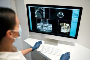
But the question of when CBCT is actually truly required – rather than simply useful or interesting – is one that the industry is still working through. And the answer, demonstrated in the IRMER principles that govern dental radiography in the UK, may have more nuance than it seems.
The justification principle in three dimensions
Every instance of radiographic exposure in UK dental practice must be justified: the expected clinical benefit must outweigh the radiation delivered. For CBCT, the dose delivered is substantially higher than for conventional 2D radiography. It varies but is typically several times that of a full-mouth periapical series, and much higher than a single intraoral radiograph.[i] That dose differential does not make CBCT unjustifiable, but it does introduce an extra layer of complexity to the choice.
Current guidance from CGDent is that CBCT should not be used as a first-line investigation where conventional 2D radiography would provide sufficient information. The imaging modality should be selected based on need, not convenience, curiosity, or the availability of the equipment.[ii] Taking a CBCT to assess a straightforward periapical area that a well-positioned periapical radiograph would adequately evaluate is likely not justifiable exposure, for example.
Where a CBCT can add real value
Where CBCT moves from useful to necessary is generally scenarios where three-dimensional information will affect the diagnosis or treatment planning in a way that two-dimensional imaging cannot.
Dental implant planning is the most commonly cited. Assessment of bone volume, bone quality, and anatomical relationships in three dimensions changes what can be planned and what can be placed with confidence. Complex endodontic cases with calcified canals, suspected vertical root fractures, or resorption, will benefit from the three-dimensional view in ways that periapical radiographs cannot replicate.[iii] Orthodontic cases involving impacted teeth are also well-established in terms of indicating CBCT necessity.[iv]
Field of view and dose optimisation
For practices with CBCT capability, the field of view selection decision is a further expression of the optimisation principle. A large field of view delivers a higher dose than a small one, and captures more, but the case itself should determine which field is appropriate. A targeted small field of view for a single-tooth endodontic assessment delivers a fraction of the dose of a large field selected out of habit or convenience.
This is a practical clinical skill that CBCT training should address directly: learning to select the minimum field of view that answers the clinical question, rather than defaulting to larger volumes on the assumption that more data is always better.
Integrating CBCT into a practice workflow
When it comes to potentially incorporating CBCT into the practice, it’s just as important to take workflow and referral into consideration. Which cases will be imaged in-house? Which will be referred? Who will report the images, and what training is in place to ensure findings both expected and unexpected are properly acted upon? A CBCT taken for an implant assessment may incidentally reveal something outside of the immediate or expected anatomical vicinity. There is still an obligation to act on this finding.

The Carestream Dental CS 9600 is a versatile 5-in-1 extraoral imaging system combining 2D panoramic, cephalometric, CBCT, 3D facial scanning, and 3D model scanning in a single compact unit. With 14 selectable fields of view from 4 x 4cm to 16 x 17cm, low-dose imaging modes, and AI-assisted patient positioning, it is designed to support the kind of clinically justified, dose-optimised CBCT that current guidance requires. Integration with CS Imaging 8 and CS 3D Imaging Premium supports reporting, planning, and guided workflows within a single environment.
The question of when to use CBCT is complex, current, and a good one to be asking. It’s important, while answering it on a case-by-case basis, to ensure that the available technology is working in harmony with the rest of the practice.

For more information on Carestream Dental visit www.carestreamdental.co.uk
For the latest news and updates, follow us on Facebook and Instagram @carestreamdental.uk
Author: Nimisha Nariapara – Trade Marketing Manager at Carestream Dental covering the UK, Middle East, Nordics, South Africa, Russia and CIS regions

[i] Li G. Patient radiation dose and protection from cone-beam computed tomography. Imaging Sci Dent. 2013;43(2):63-69. doi:10.5624/isd.2013.43.2.63
[ii] Dentistry C of G. College of General Dentistry endorses FGDP(UK) and PHE revised guidance on radiation protection. https://cgdent.uk/2020/10/13/cgdent-endorses-fgdpuk-and-phe-revised-guidance-on-radiation-protection/
[iii] Scarfe WC, Levin MD, Gane D, Farman AG. Use of cone beam computed tomography in endodontics. Int J Dent. 2009;2009:634567. doi:10.1155/2009/634567
[iv] Kapila SD, Nervina JM. CBCT in orthodontics: assessment of treatment outcomes and indications for its use. Dentomaxillofac Radiol. 2015;44(1):20140282. doi:10.1259/dmfr.20140282


