





Dr Martin Craven presents an interesting case in which conventional delayed dental implant treatment was provided to transform a patient’s failing smile.
Introduction
Comprehensive diagnostic and planning processes help to ensure the desired outcome is achieved and the patient’s expectations are met. Prosthetically-driven planning utilises 3D radiographs and intraoral scans to optimise implant and prosthetic positioning and, therefore, function, biology and aesthetics. Additionally, and importantly, digital planning helps to improve communication between patient, dentist, and dental technician, as it provides a visual presentation of the anatomical situation and the planned outcome. This is essential for managing patient expectations.[i]
Patient background
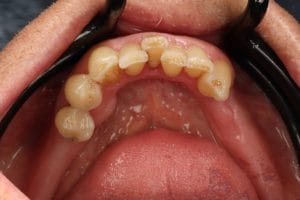
A 70-year-old male patient presented with multiple broken teeth and retained roots, seeking full-arch dental implant treatment that he had read about online.
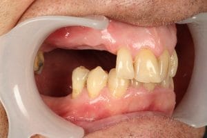
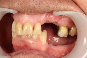
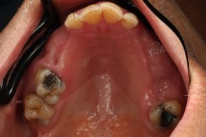
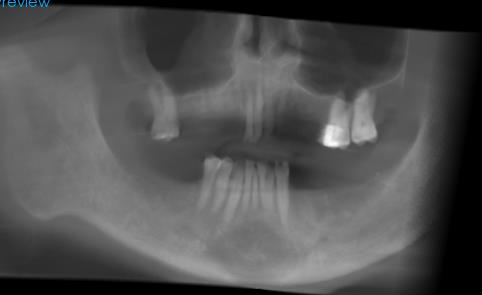
Upon assessment, the lower left and lower right molar units were missing, the LR7 region had lost the space to restore due to the over-eruption of the opposing tooth, and there were multiple retained roots which could not be restored due to lack of adequate ferrule, clinically. The patient had generalised periodontitis (stage III grade B) which was in remission at the time of presentation. Initially, a set of full mouth peri-apical radiographs and a digital iTero scan were completed, and six-point periodontal charting and bleeding scores recorded.
Initial treatment options
All treatment options were discussed with the patient, including no treatment. The unrestorable teeth and the retained roots required removal, and provisional acrylic dentures were recommended during a 12-week healing period. Long term treatment options included acrylic or cobalt-chromium partial dentures, or dental implants to restore the missing units.
Initially, the patient had requested that all of his dentition be replaced with implant supported prostheses. However, following discussion, a more conservative approach was favoured, preserving some of the teeth with a good prognosis.
A CBCT scan of both jaws would be required at 10 weeks following extraction. The patient was happy to proceed.
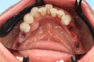
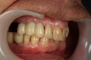
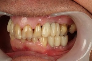
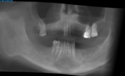
Extractions and implant planning
Immediate acrylic partial dentures (Dentuvo Silver) were produced by Ceroplast Dental Lab based on the pre-operative scans following a conformative occlusal approach.
Nine decayed teeth and retained roots were extracted in a single appointment under local anaesthetic:
- UR6 required sectioning and was removed using a closed approach
- UR5 required a simple envelope incision due to lack of visibility
- Pus was present when the UR3, 4, and 5 were removed
- Granulation tissue was present when the LL4 and 5 were removed
- No buccal bone was present around the UL5 when the socket was examined
To facilitate healing, sockets with pus and granulation tissue were debrided, and immediate dentures with socket fittings were provided. The patient was advised to wear them daily during healing, and warm salty mouthwashes were advised four times per day for seven days, beginning 24 hours post-extraction.
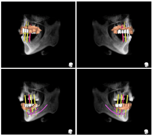
After 10 weeks of healing, a CBCT scan was requested and digital plans were created using SmileFast Dental Laboratory and RealGuide software. The designs along with the iTero scan and clinical photographs were used to explain the implant options to the patient:
- A shortened dental arch, restoring up to the premolar units only due to lack of bony height in relation to the maxillary sinus regions
- Sinus augmentation and placement of implants in the upper right molar region
- The use of implant retained bridges to facilitate the restoration of the upper right molar without the need for sinus augmentation
The latter option was selected, with a delayed loading approach recommended. Verbal agreement was recorded, before a written letter with the surgical designs was shared in order to visually display the planned treatment and aid understanding. An itemised costing was also provided with possible variations including bone grafting.
Implant treatment provision
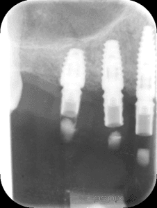
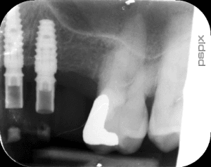
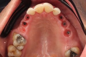
Implant surgery commenced in the upper arch. Pilot guides were used to place implants as per the digital plan. CONELOG® Progressive-Line implants were placed in the UR3, 4, 5, and UL3 and 4 positions. The UR4, 5, and UL4 implants were all very close to the sinus floor. Bone quality was D3, and standard osteotomy protocols were employed.
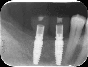
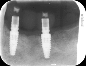
In the lower arch, CONELOG® Progressive-Line implants were placed in the LR6 and 7, the LL4 and 6, using a pilot guide. An envelope incision made to visualise the LL4 region revealed insufficient buccal bone to place the implant. This possibility had already been outlined, and was discussed with the patient during surgery. The patient consented to an allograft bone augmentation and the implant was placed using the guide. The exposed buccal threads were grafted using MinerOss® Blend, and a porcine-derived collagen xenograft membrane (Mem-Lok® Pliable) was used to secure the graft. Amoxicillin was provided to reduce the risk of infection, and care was taken to fully cover the membrane, using simple interrupted 4-0 Vicryl Rapide sutures.
The patient was reviewed twice in a two-week period to monitor healing due to the higher risk of infection. The implants were fully submerged, with a cover screw placed and hand tightened. The patient was provided with a Curaprox implant care kit, which included an Ultra Soft toothbrush, and a 0.2% aqueous chlorhexidine mouthwash to be used four-times per day for 10 days following surgery.
Six weeks post-operative radiographs were taken to assess bone levels and potential pathology around the implant sites. At four months post-op, the implants were uncovered and healing caps were placed for a further two weeks.
An iTero scan was taken, and SmileFast Dental Lab provided provisional bridges and crowns to restore the dental implants, using PMMA on Ti stock abutments. Radiographs confirmed adequate bone levels at the time of loading.
Two weeks later, an issue occurred with the provisional restorations. The UR6 pontic fractured due to overloading of the PMMA material. The patient understood that this was less likely with the definitive bridges. During the provisional stage, the patient requested tooth whitening, for which we provided 10% carbamide peroxide in close fitting custom trays over a two-week period.
After three months of provisionalisation, the implants were loaded with definitive restorations – monolithic zirconia and custom Ti abutments from SmileFast Dental Lab. Minimal edge bonding of the upper anterior teeth was also completed.
Outcome and case appraisal
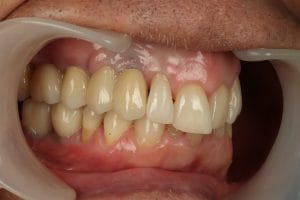
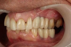
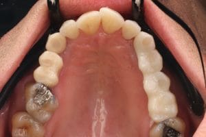
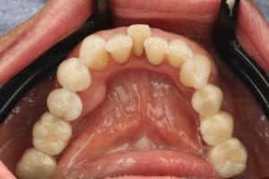
The patient and I have been very pleased with the results, achieving the patient’s primary aim of good function whilst eating. The patient’s home care and knowledge of oral hygiene has improved during the process, and he is caring well for his implants and natural teeth.
The digital workflow allowed greater consideration of how to navigate potential issues such as the poor bone quality in the UR6 region. The pilot guides allowed reasonable precision in delivering the treatment plans, whilst also allowing some degree of flexibility – this can be useful when there are occasional changes required, such as the need to perform augmentation at the LL4. On reflection, I believe this case would have been equally well-suited to a fully guided workflow. Planning the case with a staged approach meant that the bone quality was optimised for implant placement, and an overall good thickness of keratinised tissue around the implants was achieved using provisional restorations.
I would highly recommend introducing digital planning to any implant cases. Many clinicians seem to suggest digital planning and guides are reserved for the most complex cases only. However, I find that even a single implant placed between two teeth can greatly benefit from a digital plan and guide – achieving optimal position in the bone. In my opinion, this offers greater predictability of long-term success. I also find digital plans to be a great tool facilitating patient trust, allowing patients to see the level of detail and consideration put into the planning.
For product information from BioHorizons and Camlog, please visit https://theimplanthub.com/
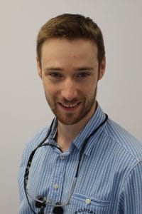 Author bio: Dr Martin Craven BDS BSc(Hons) PGDip(implant dentistry) MFDS RCS Ed graduated in 2013 from Peninsula Dental School. His early career was spent in Oral & Maxillofacial Surgery at Peterborough Hospital, becoming a principal of a private practice in 2018 based in Redditch, the West Midlands. In 2022-2025, he completed a masters level diploma in implant dentistry with the BioHorizons education programme, and has also completed further education on guided surgery with Dr Nick Fahey.
[i] Schubert, O., Schweiger, J., Stimmelmayr, M., Nold, E. and Güth, J.F., 2019. Digital implant planning and guided implant surgery–workflow and reliability. British dental journal, 226(2), pp.101-108.


