






The panoramic radiograph is one of the most familiar images in dentistry. Practices use them routinely for assessments, treatment planning, orthodontics and implantology. They are generally taken quickly, interpreted quickly, and filed away.
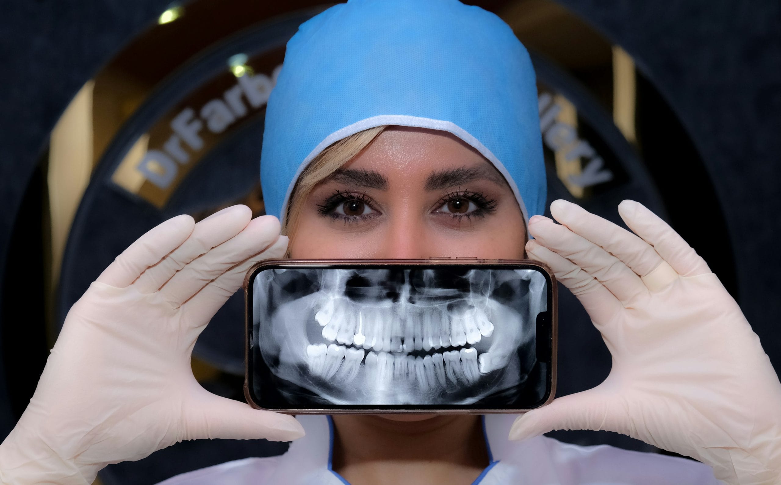
But there is a growing body of clinical evidence strongly suggesting that clinicians who treat the panoramic radiograph as a strictly dental document may be leaving potentially important information unread.[i]
Research consistently shows that a meaningful proportion of routine panoramic radiographs contain findings entirely unrelated to the primary dental indication. A study analysing over 2,000 panoramic radiographs found that 26% contained incidental findings beyond caries, periodontal, and periapical disease.[ii] These included carotid artery calcifications, maxillary sinus pathology, tonsilloliths, cysts, and bone lesions. More than a quarter of images, in other words, contained information that could be clinically relevant –and that could easily go unnoticed if the clinician’s focus is solely on the dentition.
Beyond the teeth and jaw
The most clinically significant of these incidental findings, and the one attracting the greatest research attention, is carotid artery calcification (CAC). The carotid region is frequently captured in the field of a panoramic radiograph, and calcified atheromatous plaques can appear as radiopaque nodular masses adjacent to the cervical vertebrae at the level of C3 and C4. Studies have found CAC in up to 38% of panoramic radiographs where the carotid region is adequately visualised, with prevalence increasing with age and in patients with known cardiovascular risk factors.[iii]
The implications of this are not trivial by any means: CAC detected on panoramic radiographs has been associated with elevated rates of cerebrovascular accident, coronary artery disease, hypertension, and hyperlipidaemia. One study examining 314 patients with radiographically identified CAC found a stroke prevalence of 15% and coronary artery disease in 28% of that group.[iv] A 2026 study further found that specific features of CAC on panoramic imaging correlated with more severe vascular disease on carotid ultrasound.[v]
It is important to be clear about the limits of these findings, however: the panoramic radiograph is not a vascular screening tool, and its sensitivity for CAC detection is imperfect. A clinician who identifies a suspicious radiopacity in the carotid region should refer appropriately for further investigation without attempting to draw definitive conclusions. Nevertheless, the evidence supports a clear professional responsibility: when an image is in the patient’s record, every structure visible within it warrants some consideration.
Sinus, bone, and soft tissue
Further to vascular findings, the panoramic radiograph offers a view of other structures easy to overlook in a quick appointment. Sinusitis in the maxillary sinus, for example, is visible on a panoramic image and may require onward referral to a medical colleague. Elongated styloid processes, found in up to 33% of panoramic radiographs in some studies,[vi] can be associated with Eagle’s syndrome and orofacial pain that patients may have attributed to dental causes. Bone lesions, including cysts and radiolucent areas beyond the periapical region, may require further investigation, too.
The point here is to simply recognise that a panoramic radiograph, taken for routine appointments, does capture a wider field than just the teeth and jaw. A more thorough interpretation of this data is an acknowledgement of this fact.
How much does image quality matter?
A clinician cannot act on a finding they cannot see clearly. The diagnostic value of a panoramic radiograph is fundamentally dependent on the clarity and consistency of the image produced. Poor positioning, artefacts, or inconsistent image quality will all reduce the likelihood that incidental findings will be identified, as well as increase the probability of retakes, which will subject the patient to extra exposure.
This is where investment in solid imaging equipment becomes a question of safety. The Carestream Dental CS 8200 3D Access is a compact 4-in-1 system combining panoramic imaging, CBCT, 3D model scanning, and optional cephalometric capability – designed to bring advanced imaging within reach of general dental practices without sacrificing ease of use. Panoramic programmes include segmented bitewing, TMJ, and maxillary sinus views, ensuring that the full field captured in every image is supported clinically. CS MAR technology reduces metal artefacts for more accurate interpretation, a low-dose mode is available for both 2D and 3D examinations, and 75-micron CBCT resolution supports precise diagnosis across endodontic and implant indications when 3D imaging is required. The compact footprint and intuitive interface make it straightforward to integrate into practices of any size.
The panoramic radiograph has always offered more than a view of the dentition. Recognising that, and ensuring that the images produced are consistently clear enough to act on, is a straightforward way to deliver more comprehensive care to every patient in the chair.

For more information on Carestream Dental visit www.carestreamdental.co.uk
For the latest news and updates, follow us on Facebook and Instagram @carestreamdental.uk
Author: Nimisha Nariapara – Trade Marketing Manager at Carestream Dental covering the UK, Middle East, Nordics, South Africa, Russia and CIS regions.

[i] Almeida FT, Gianoni-Capenakas S, Rabie H, Figueiredo R, Pacheco-Pereira C. The use of panoramic radiographs to address the oral health needs of vulnerable Canadian populations. Can J Dent Hyg. 2024;58(1):19-25. Published 2024 Feb 1.
[ii] Ghassemzadeh S, Sbricoli L, Frigo AC, Bacci C. Incidental findings detected with panoramic radiography: prevalence calculated on a sample of 2017 cases treated at a major Italian trauma and cancer centre. Oral Radiol. 2021;37(3):507-517. doi:10.1007/s11282-020-00488-1
[iii] Alves N, Deana NF, Garay I. Detection of common carotid artery calcifications on panoramic radiographs: prevalence and reliability. Int J Clin Exp Med. 2014;7(8):1931-1939. Published 2014 Aug 15.
[iv] Brar A, DeColibus K, Rasner DS, et al. Carotid Artery Calcification Detected on Panoramic Radiography Is Significantly Related to Cerebrovascular Accident, Coronary Artery Disease, and Poor Oral Health: A Retrospective Cross-Sectional Study. Dent J (Basel). 2024;12(4):99. Published 2024 Apr 10. doi:10.3390/dj12040099
[v] Karlsson A, Gustafsson N, Wester P, Zamure-Damberga L, Levring Jäghagen E. Carotid artery calcifications on panoramic radiographs are associated with vascular disease severity on carotid ultrasound. Dentomaxillofac Radiol. 2026;55(1):28-36. doi:10.1093/dmfr/twaf061
[vi] Bruno G, De Stefani A, Balasso P, Mazzoleni S, Gracco A. Elongated styloid process: An epidemiological study on digital panoramic radiographs. J Clin Exp Dent. 2017;9(12):e1446-e1452. Published 2017 Dec 1. doi:10.4317/jced.54370


