






In a bid to make complex treatments more predictable, dental imaging has become a key source of information for all dental professionals. In endodontic care, an understanding of each patient’s unique anatomy and canal structure is vital. Without it, clinicians could injure patients or deliver ineffective care that requires retreatment.

Clinicians need to understand how advanced imaging software and systems should be chosen for effective endodontic results today, as well as anticipate common problems in order to effectively minimise the need for repeated exposures.
Imaging needs in endodontics
In endodontic care, the quality and availability of dental imaging is vital for the diagnosis and treatment of an issue. Cone beam computed tomography (CBCT) creates 3D images which can help determine the aetiology of an endodontic problem. This offers clinically relevant information to a clinician, which leads to better treatment planning decisions and more predictable results.[i]
They have become an essential and widespread solution. A survey of 1083 endodontists in the US, carried out in 2017, found that 80.30% of respondents had access to a CBCT scanner.[ii] This includes on-site and off-site solutions. The most common uses were for internal or external resorptions, preoperatively for surgical retreatment or intentional replantation, and analysing missing canals.ii Indications for use in endodontic treatment also include evaluating intracanal foreign materials (such as a separated instrument), to identify and assess the extent of apical periodontitis, and for the healing assessment or follow-up treatment.[iii]
Tasks such as analysing the number of canals, their positioning and their curvature can also be completed with a CBCT solution.
Clinicians can refer patients out of the practice for the imaging needs, or treat them completely in-house with the correct system (and attract referrals from local practices too). Finding a CBCT scanner that will reliably deliver high-quality results, with minimal need for retakes is key.
Aware of the risks
Misuse of CBCT equipment can not only waste precious clinical time and expense, but also put patients at risk of harm. Staff should have their training updated after a CBCT scanner is introduced to the practice.[iv] This includes those that may not work directly with radiography; members of the dental team should be able to recognise the significance of warning lights and signs around a designated room for CBCT imaging, and the restrictions placed on access to this space when in use.iv
CBCT systems use elements of traditional radiography to produce an image. This requires the exposure of a patient to X-rays. We are all exposed to radiation every day, but a typical CBCT scan of the jaw makes a patient subject to the equivalent of 6 to 30 days of background exposure; thought this is lower than traditional CT scans of the jaw, which produces radiation similar to 63 to 154 days of exposure.[v]
A 2021 study found that repeat use of CBCT scans at the maximum exposure settings could increase the risk of cancer development, with higher hazards for women and children.[vi] Repeated use over prolonged periods of time (as potentially seen in orthodontic treatment) is not typically expected in endodontic care, and justified scans will ensure patients are protected.
Be aware that the need for repeated CBCT scans can be born out of clinical error. For example, CBCT scans can create metal artefacts in an image if a nearby restoration uses a material such as amalgam. Results that include an artefact can limit virtual planning and the accuracy of guided endodontic techniques, which help create more accurate interventions.[vii] If a clinician does not limit their field of view accordingly, the tooth destined for treatment could be obscured, and another scan is necessary for safe and effective care.
Finding your solution
When choosing a high-quality CBCT system for your practice, there is so much to consider. It must be accurate, with the ability to present clear images of the dentition. Versatility is also useful, with different fields of view allowing clinicians to minimise the risk of metal artefact creation whilst limiting exposure – advanced solutions with metal artefact reduction can aid treatment, even when such an error occurs.
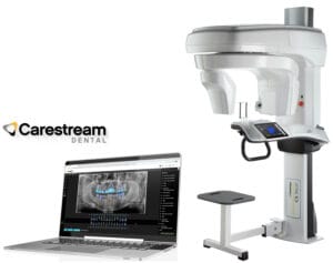
This is all found, alongside much more, in the CS 9600 CBCT Scanner from Carestream Dental. The scalable, 5-in-1 solution delivers smart innovations at every exam. 14 different fields of view improve the abilities of clinicians to gain an accurate, safe result from each scan, whilst a low-dose imaging mode reduces X-ray exposure – meaning 3D images can be attained at up to an 86% lower dose than 2D panoramic images. CS MAR (metal artefact reduction) also reduces the influence of artefacts, whilst allowing dynamic image comparison for complete confidence.
The CBCT scanner has become an essential tool for many endodontists. With complete understanding of its abilities, and safe use, clinicians can help even more patients by forming predictable treatment plans – it just starts with a scan.

For more information on Carestream Dental visit www.carestreamdental.co.uk
For the latest news and updates, follow us on Facebook and Instagram @carestreamdental.uk
 Author: Nimisha Nariapara – Trade Marketing Manager at Carestream Dental covering the UK, Middle East, Nordics, South Africa, Russia and CIS regions.
[i] Boquete-Castro, A., Lopez, A. P., Martins, A. S., Lorenzo, A. S., & Perez, P. R. (2022). Applications and advantages of the use of cone-beam computed tomography in endodontics: An updated literature review. Saudi Endodontic Journal, 12(2), 168-174.
[ii] Setzer, F. C., Hinckley, N., Kohli, M. R., & Karabucak, B. (2017). A survey of cone-beam computed tomographic use among endodontic practitioners in the United States. Journal of endodontics, 43(5), 699-704.
[iii] Setzer, F. C., & Lee, S. M. (2021). Radiology in endodontics. Dental Clinics, 65(3), 475-486.
[iv] Health Protection Agency, (2010). Guidance on the Safe Use of Dental Cone Beam CT (Computed Tomography) Equipment. (Online) Available at: https://assets.publishing.service.gov.uk/media/5a7d4f13ed915d321c2de451/HPA-CRCE-010_for_website.pdf [Accessed August 2025]
[v] NHS Guy’s and St Thomas’ NHS Foundation Trust, (2024). Dental cone beam CT scan (CBCT). (Online) Available at: https://www.guysandstthomas.nhs.uk/health-information/dental-cone-beam-ct-scan-cbct [Accessed August 2025]
[vi] Jha, N., Kim, Y. J., Lee, Y., Lee, J. Y., Lee, W. J., & Sung, S. J. (2021). Projected lifetime cancer risk from cone-beam computed tomography for orthodontic treatment. Korean journal of orthodontics, 51(3), 189-198.
[vii] Decurcio, D. A., Bueno, M. R., Silva, J. A., Loureiro, M. A. Z., Sousa-Neto, M. D., & Estrela, C. (2021). Digital planning on guided endodontics technology. Brazilian dental journal, 32, 23-33.


