






External cervical resorption (ECR) is an often underdiagnosed condition in which the tooth undergoes pathological loss of hard tissue, starting at the cervical region, just apical to the epithelial attachment. Its presentation can be subtle, with general dental practitioners (GDPs) usually the first to encounter suspicious radiographic or clinical signs, making their role in early detection and referral essential. This article explores the nature, challenges, and management of ECR, using a referral from a GDP to a UK dental hospital as an example of good practice.
The nature of external cervical resorption
A sub-type of external root resorption, ECR is a localised resorptive defect near the cervical margin.[i] Unlike caries, it is not primarily microbial in origin but is associated with damage, loss or deficiency of the periodontal ligament and subepithelial cementum, which renders the root surface vulnerable to odontoclastic activity. Once initiated, odontoclasts and multinucleated giant cells resorb dental hard tissues, creating irregular resorptive lacunae. Importantly, the pulp typically remains unaffected in early lesions due to the protective layer of pre-dentine, meaning the process is usually asymptomatic until advanced stages.[ii]
ECR may manifest clinically or, more often, be detected incidentally on radiographs. The exact aetiology is multifactorial with risk factors including orthodontic treatment, trauma, domestic cat ownership, parafunctional habits, intracoronal bleaching, periodontal therapy, and surgical procedures. In many cases, however, no clear cause is identified.[iii]
Classification and management principles
Heithersay’s grading system is based on 2D peri-apical radiographs.ii The system divides ECR lesions into four classes, depending on the size and extent of the resorptive process into the dentine. Early lesions are often amenable to conservative treatment, whereas advanced lesions may necessitate extraction. However, bi-dimensional interpretation does not provide accurate assessment and should not be relied upon to establish treatment plans.
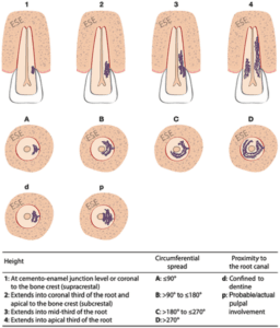
By contrast, the Patel classification (2018)[iv] is a CBCT-based system that helps in assessing the extent and location of the resorptive defect. The classification has three key components – the height of the lesion (coronal-apical extent), circumferential spread, and proximity to the pulp (Figure 1).
At this stage, it is pertinent to note that cases of ECR often affect multiple teeth, which should influence our assessment clinically and radiographically.
Management strategies should ensure that a tooth diagnosed with ECR is maintained in a healthy and functional status, avoiding tooth extraction and improving aesthetic outcomes. To arrest the resorptive process, affected tissue requires excavation and the remaining defect requires closure with an effective coronal seal/restoration provided.[v]
Two main therapeutic strategies can be proposed:
- Internal repair – small lesions with inaccessible points of entry can only be treated via endodontic treatment and restoration of the lesion.
- External repair – accessing surgically when the lesion extends farther apically than the gingival margin. Resorptive tissue is removed, and the defect is restored.[vi]
Endodontic involvement – If the lesion has progressed close to or into the pulp, root canal treatment may be indicated in combination with defect restoration.
Alternative options can include:
- Non-surgical monitoring – In some early or inaccessible cases, periodic radiographic review may be appropriate, especially in older patients or where intervention carries greater risk.
- Extraction – For extensive lesions, especially Heithersay Class 4, prognosis is poor, and extraction is often the most appropriate option.
Early detection: The GDP’s role
Early diagnosis of ECR is challenging. For GDPs, vigilance during routine clinical examinations and radiographic assessments is key.
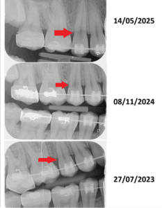
The referral below provides an exemplary model whereby the GDP identified subtle but concerning radiographic changes around the cervical aspect of a tooth and promptly referred the patient for specialist assessment (Figure 2).
Clinically, suspicious findings include:
- A pinkish cervical hue, indicating granulation tissue beneath enamel
- Unusual surface textures detected with probing
- Unexplained radiolucencies near the cervical region
Radiographically, ECR may appear as an irregular radiolucency superimposed on the root canal space. Traditional two-dimensional radiographs are limited, as the defect may be masked or misinterpreted as caries or cervical burnout. Increasingly, CBCT is regarded as the gold standard for accurate diagnosis, as it allows three-dimensional assessment.vi However, CBCT is not typically available in general dental practices, further emphasising the GDP’s role in timely referral.
GDPs should adopt a low threshold for suspicion when faced with signs, especially in patients with known risk factors.
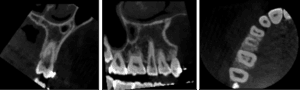
The example referral facilitated access to specialist imaging and classification of the lesion, allowing a tailored treatment plan to be developed (Figure 3). Without this timely referral, the lesion may have progressed unnoticed, severely compromising the tooth’s prognosis.v
Reflections on the referral example
The referral demonstrates key qualities of best practice for GDPs:
- Recognition of abnormal findings
- Clear communication in the referral letter
- Patient advocacy
This approach embodies the collaborative relationship between primary and secondary care, optimising patient outcomes in more complex dental conditions such as ECR.
The British Endodontic Society (BES) is open to anyone with an interest in endodontics, including early career dentists, GDPs and experienced practitioners, as well as specialists. BES members gain access to high-quality education and support, as well as networking opportunities, to ensure they stay abreast of the latest developments in the field.
Conclusion
ECR is a challenging condition to detect and manage, owing to its asymptomatic nature and subtle early presentation. GDPs play a critical role in identifying suspicious features and ensuring timely referral for advanced investigation and treatment.

For more information about the BES, or to join, please visit the website www.britishendodonticsociety.org.uk or call 07762945847
 Author Bio: Roshni Ruparelia is currently a Specialty Registrar in Restorative Dentistry at Leeds Dental Institute, pursuing her keen interest in endodontics. She has been recognised for her clinical case presentations and contributes actively to the field as a committee member of the British Endodontic Society’s Early Careers Group. As well as dentistry, Roshni holds a BSc in Psychology, allowing her to bring a unique interdisciplinary perspective to patient care and professional development.
[i] Patel, S. and Ford, T.P., 2007. Is the resorption external or internal? Dental update, 34(4), pp.218-229.
[ii] Heithersay, G. S. (1999). Clinical, radiologic, and histopathologic features of invasive cervical resorption. Quintessence International, 30(1), 27–37.
[iii] Patel, S., Abella, F., Patel, K., Bakhsh, A., Lambrechts, P. & Al-Nuaimi, N., 2025. Potential predisposing features of external cervical resorption: An observational study. International Endodontic Journal, 58(5), pp.799–801. https://doi.org/10.1111/iej.14216
[iv] Patel S, Foschi F, Mannocci F, Patel K. External cervical resorption: a three-dimensional classification. Int Endod J. 2018 Feb;51(2):206-214. doi: 10.1111/iej.12824. Epub 2017 Sep 13. PMID: 28746776.
[v] Talpos-Niculescu RM, Nica LM, Popa M, Talpos-Niculescu S, Rusu LC. External cervical resorption: Radiological diagnosis and literature (Review). Exp Ther Med. 2021 Oct;22(4):1065. doi: 10.3892/etm.2021.10499. Epub 2021 Jul 27. PMID: 34434279; PMCID: PMC8353645.
[vi] Patel, S., Mavridou, A., Lambrechts, P., & Saberi, N. (2019). External cervical resorption-part 1: histopathology, distribution and presentation. International Endodontic Journal, 51(11), 1205–1223.


