






Accurate dental impressions are fundamental to successful treatment planning, precise restoration manufacture, and predictable clinical outcomes. Selecting the most appropriate method for capturing this data helps minimise inaccuracies and gives clinicians confidence that the care they provide will deliver reliable, long-lasting results for their patients.
Digital technologies now play an important role in modern dentistry, and intraoral scanners (IOS) are firmly established in many practices. However, as with any clinical tool, they are not designed to address every scenario. Understanding where different impression methods perform best allows clinicians to make informed, case-specific decisions.
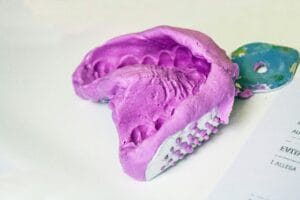
Where intraoral scanners work well
Intraoral scanners are highly effective in a range of routine indications. They are commonly used for single-unit restorations, short-span bridges, and smaller quadrant-based prostheses, where scan paths are limited and stitching requirements are minimal.[i] In these cases, IOS can offer speed, convenience, and seamless integration into digital workflows.
IOS are also widely used in orthodontic treatment, supporting digital planning and aligner workflows. As with all impression techniques, outcomes depend on case complexity, patient anatomy, and operator experience, and accuracy should always be evaluated on an individual basis.[ii]
Understanding the limits of any single approach
Not all clinical situations are equally suited to intraoral scanning. Certain factors can make reliable data capture more challenging, particularly when accuracy across larger or more complex areas is required.
Moisture control is one such consideration. Saliva, blood, and gingival fluid can interfere with data capture, and maintaining a consistently dry field is not always achievable. Similarly, deep subgingival margins present a challenge, as scanners can only record what is directly visible.[iii]
Case size also plays an important role. Full-arch and long-span scans require the stitching together of multiple images, increasing the potential for cumulative distortion. Operator technique and experience further influence outcomes, particularly in complex cases.[iv]
These considerations do not diminish the value of intraoral scanners, but they do highlight that no single impression method is optimal for every clinical indication.
When conventional impressions matter
For larger and more complex restorations, conventional impression techniques continue to play an important role in achieving predictable outcomes. This is particularly true for full-arch restorations, implant-supported work, and complete dentures, where accurately capturing soft-tissue anatomy, functional extension, and border detail is critical. In particular, when taking impressions in complete denture cases, it is essential to use a mucocompressive technique – applying controlled pressure to the oral tissues – to maximise stability when the patient is chewing. This is an example of a requirement which digital impressions cannot replicate, and where the retention of final dentures would, therefore, be compromised.
Clinical research supports this distinction. A controlled crossover study[v] by Chebib et al. (Fit and retention of complete denture bases: Part II – conventional impressions versus digital scans) demonstrated that complete denture bases produced from conventional impressions showed superior retention compared to those produced from digital scans. This finding underlines the continued relevance of conventional techniques in situations where mucosal support and functional adaptation are key to success.
Conventional impressions can also offer greater reliability when deep margins are present, moisture control is compromised, or long-span accuracy is required.iv In these situations, the physical properties of impression materials can provide a more complete and stable representation of the clinical environment.
Importantly, choosing a conventional impression does not mean stepping away from digital dentistry. Rather, it reflects a considered decision to prioritise accuracy and predictability for the specific case.
Bringing conventional impressions into the digital workflow
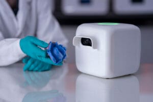 The Cubit360™ scanner from Mimetrik™ enables conventional impressions to be fully integrated into modern digital workflows. As the world’s only six degrees of freedom scanner, Cubit360™ is designed specifically to digitise conventional dental impressions with exceptional detail and consistency.
The system allows fast and intuitive scan capture, making it particularly well suited to complex treatment types where accuracy is critical. By simply positioning the impression in front of the scanner, a high-quality digital file can be created and shared with the laboratory immediately. This removes the risks associated with physical transport, such as distortion, loss, or damage.
Choosing the right tool for each case
Intraoral scanners play an important role in contemporary dentistry and are an excellent choice for many indications. However, not all cases are ideally suited to intraoral capture. For situations where conventional impressions offer greater accuracy, the ability to digitise those impressions efficiently and reliably becomes invaluable.
By combining established impression techniques with advanced digitisation through Cubit360™, clinicians can maintain accuracy across a broader range of cases—without sacrificing the efficiency, connectivity, and convenience of digital workflows.
Making informed, case-specific decisions about impression taking ultimately supports better clinical outcomes and greater confidence for both clinicians and patients.

For more information about Mimetrik, please visit https://mimetrik.tech/
 Author: Alyn Morgan
[i] Sacher, Mattia et al. “Accuracy of commercial intraoral scanners.” Journal of medical imaging (Bellingham, Wash.) vol. 8,3 (2021): 035501. doi:10.1117/1.JMI.8.3.035501
[ii] Christopoulou, Isidora et al. “Intraoral Scanners in Orthodontics: A Critical Review.” International journal of environmental research and public health vol. 19,3 1407. 27 Jan. 2022, doi:10.3390/ijerph19031407
[iii] Camcı, H. and Salmanpour, F., 2020. Effect of saliva isolation and intraoral light levels on performance of intraoral scanners. American Journal of Orthodontics and Dentofacial Orthopedics, 158(5), pp.759-766.
[iv] Mourouzis, P., 2025. Critical methodological factors influencing the accuracy of intraoral scanners in digital dentistry research. Computers in Biology and Medicine, 187, p.109780.
[v] Chebib, N., Imamura, Y., El Osta, N., Srinivasan, M., Müller, F. and Maniewicz, S., 2024. Fit and retention of complete denture bases: Part II–conventional impressions versus digital scans: A clinical controlled crossover study. The Journal of Prosthetic Dentistry, 131(4), pp.618-625.


