






 Nimisha Nariapara – Trade Marketing Manager at Carestream Dental covering the UK, Middle East, Nordics, South Africa, Russia and CIS regions
Amalgam fillings, silver and golden crowns, and the implants supporting aesthetic restorations are all examples of metals used in dental care. Silver amalgam, in particular, is thought to be the most common material for NHS permanent fillings across the UK,[i] and 80% of adults in the UK report to have fillings (though not all of these will be amalgam).[ii]
Each of these can help restore function to a patient’s dentition, minimising pain from previous injuries or caries-affected teeth – but they’re not free from problems. In particular, clinicians can run into difficulties when assessing radiographs, and encountering metal artefacts.
Dental professionals should understand why metal artefacts are created during imaging routines, and be aware of the ways they can be minimised to improve diagnosis and treatment planning.
Impact on imaging
An image artefact is a feature or discrepancy produced in a radiograph that is not present in real life.[iii] In dental care, artefacts can interfere with clinical assessments or treatment plans, leading to inaction or overtreatment, with patients in turn receiving compromised care. Metal features in particular are an issue, as false dark and bright regions and linear streaks can appear close to or radiating from the object in a radiograph.iii
When a clinician uses a cone beam computed tomography (CBCT) system, X-ray beams pass through dental tissue, which alters the energy spectrum of the beam. These differences produce the final radiograph that allows clinicians to assess hard and soft tissues, and additional structures. Metallic objects are known to alter this energy the most, and can influence image quality by reducing contrast and obscuring the nearby structures.[iv]
In some cases, metals can be avoided from a radiographic scan entirely. A clinician may only need to analyse the posterior dentition, but a patient has a metal implant placed in the anterior incisal region, for example. The correct selection of a limited field of view (FOV) that localises the scan would omit the metal item, and the artefacts it would produce. This would also be beneficial as it would follow the ‘as low as reasonably possible’ (ALARP) approach to radiography, which limits exposure to minimise the possibility of adverse health effects.
However, treatment needs don’t always work this way. A metal object could be immediately adjacent to the area of interest, or otherwise entirely unavoidable if an image of the entire dentition is required. In turn, clinicians should be prepared to use alternative systems for metal artefact reduction (MAR).
What is MAR?
There are many ways to reduce the impact of metal restorations on a radiographic image. An increase in the tube current or voltage can reduce metal artefacts by increasing the number of photons with a higher energy reaching the detector – but this creates an increased radiation exposure for the patient, an ultimately unwanted outcome considering the ALARP principle.[v]
Alternatively, MAR algorithms have been developed that help to clear up obstructions, improving visibility and the ability to create better-informed treatment plans. Deep learning models have shown promising results,[vi] and alternative algorithms are understood to reduce artefacts when applied before or after image acquisition.[vii]
It’s important to understand the need for care with MAR systems. The type and size of defect, type of CBCT system used, type of MAR algorithm used, and the experience of the radiologist are all variable factors when acquiring radiographic results for diagnosis or treatment planning.[viii] Controlling as many aspects as possible is vital to consistently creating reliable radiographs for diagnosis or treatment planning.
How MAR makes an impact
Clinicians may be able to avoid overtreatment, or incorrect diagnosis leading to retreatment, if an area of the dentition is no longer obscured by a metal restoration’s artefacts. This is only possible when using an effective MAR system from a reliable source, created and supported by names that dental professionals can trust.
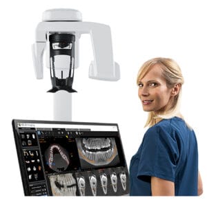 The CS 8200 3D Access is a leading CBCT system from Carestream Dental, which offers effective CS MAR technology with unique live comparisons, aiding the confirmation of diagnosis and reducing the risk of misinterpretation. The system also features six different fields of view, which allow clinicians to target select areas of the dentition to avoid artefact production. Dental professionals with limited radiographic experience will benefit with the CS 8200 3D Access as it makes advanced CBCT imaging more accessible, with a modern, user-friendly interface.
With many patients having metal-based restorations, it’s important to find ways to assess the dentition without detrimental impacts on care. MAR algorithms and targeted approaches to CBCT imaging can help clinicians attain optimal results – experience and high-quality solutions are all that is needed.

For more information on Carestream Dental visit www.carestreamdental.co.uk
For the latest news and updates, follow us on Facebook and Instagram @carestreamdental.uk
[i] British Dental Journal, (2024). Amalgam ban may hasten NHS dentistry’s demise. British Dental Journal, (Online) Available at: https://www.nature.com/articles/s41415-024-7073-9 [Accessed July 2025]
[ii] Office for Health Improvement & Disparities, (2024). Adult oral health survey 2021: self-reported health of teeth and gums. GOV.UK. (Online) Available at: https://www.gov.uk/government/statistics/adult-oral-health-survey-2021/adult-oral-health-survey-2021-self-reported-health-of-teeth-and-gums [Accessed July 2025]
[iii] Hinchy, N. V., Anderson, N. K., & Mahdian, M. (2022). Metal artifact reduction using common dental materials. Dentomaxillofacial Radiology, 51(2), 20210302.
[iv] Bechara, B. B., Moore, W. S., McMahan, C. A., & Noujeim, M. (2012). Metal artefact reduction with cone beam CT: an in vitro study. Dentomaxillofacial Radiology, 41(3), 248-253.
[v] Kleber, C. E., Karius, R., Naessens, L. E., Van Toledo, C. O., van Osch, J. A., Boomsma, M. F., … & van der Molen, A. J. (2024). Advancements in supervised deep learning for metal artifact reduction in computed tomography: A systematic review. European Journal of Radiology, 181, 111732.
[vi] Kleber, C. E., Karius, R., Naessens, L. E., Van Toledo, C. O., van Osch, J. A., Boomsma, M. F., … & van der Molen, A. J. (2024). Advancements in supervised deep learning for metal artifact reduction in computed tomography: A systematic review. European Journal of Radiology, 181, 111732.
[vii] Bohner, L., Parize, H., Cordeiro, J. V. C., Laureano, N. K., Kleinheinz, J., Caldas, R. A., & Dagassan-Berndt, D. (2025). Bone quality assessment around dental implants in cone-beam CT images: effect of rotation mode and metal artefact reduction tool. Dentomaxillofacial Radiology, 54(4), 286-293.
[viii] Salemi, F., Jamalpour, M. R., Eskandarloo, A., Tapak, L., & Rahimi, N. (2021). Efficacy of metal artifact reduction algorithm of cone-beam computed tomography for detection of fenestration and dehiscence around dental implants. Journal of biomedical physics & engineering, 11(3), 305.


