






 Author: Alyn Morgan
Imaging is a cornerstone of dental diagnosis and treatment planning, making it absolutely crucial that it be carried out correctly to achieve accurate results. However, there are a number of challenges that dental professionals may face when capturing dental images, which mean that the resulting scans are sometimes unusable and retakes are necessary. As such, it’s important to consider the causes and consequences of inaccurate images, and understand how they can be minimised to improve patient management.
Implications of movement during diagnostic imaging (CBCT)
Should a patient move during a CBCT examination, artifacts may arise. In resulting images, these may present as stripes/streaks, double contours, or overall lack of sharpness. The presence of artifacts such as this, in extreme cases, may render images uninterpretable. This means that clinicians are unable to make a diagnosis or discern relevant information based on the images. Research suggests that, whenever movement is present, image quality is hampered. Although, the risk of an image being ‘not interpretable’ becomes statistically significant only when movement is 3 mm or more.[i]
When a scan is deemed uninterpretable, a retake will be needed in order to produce an image of sufficient quality. For the patient, this means an extra dose of radiation exposure which could have been prevented if there hadn’t been movement.
How to minimise patient movement during scans
In order to effectively minimise patient movements during diagnostic scans, patient education is crucial. This includes informing patients about the risks presented by ionizing radiation (doubled with retakes), in addition to the importance of regular follow up appointments to reduce otherwise unnecessary examinations. The minimisation of repeated exposures in diagnostic imaging needs a comprehensive approach – encompassing innovative technology, optimising techniques, and educating patients. Collectively, these approaches will help ensure that benefits of diagnostic imaging are maximised whilst patients are more effectively protected from the potential risks associated with radiation exposure.[ii]
What are the considerations during intraoral scanning?
In single and partial implant restorations, research finds the use of intraoral scanners satisfactory and reproducible. However, in full arch scenarios, they remain problematic and unreliable – this is likely due to the lack of landmarks in an edentulous mouth. Full arch cases can require a long scanning process, often resulting in several interruptions, and the potential need to restart the scan from the beginning. Many modern intraoral scanners aim to limit these challenges, however, tracking loss can occur for a number of reasons. These include patient movement, the presence of reflective or transparent surfaces, saliva, and human error.[iii]
Scanning speed is also thought to play a part in scan accuracy. Elevated scanning speed may compromise accuracy, due to limited capability to capture adequate data points to achieve a complete 3D reconstruction. However, scanning at an excessively slow speed could increase the risk of patient movement. To optimise accuracy of the scan, clinicians must have a good understanding of how to use their particular scanner.[iv]
The importance of accuracy in dentistry
As mentioned, there are a number of factors which might interfere with intraoral scan accuracy, including patient compliance, mouth opening, tongue size, and movement, in addition to the need for practitioners to invest time in learning how to use the device effectively. However, it is important that accuracy is prioritised in order to support optimal diagnosis, treatment planning, and communication (with patients and colleagues).[v] As such, in cases where intraoral scanners typically encounter challenges, such as full arch edentulous cases, it may be more appropriate to take impressions using analogue methods. With modern technological solutions, traditional impressions can be digitised with complete accuracy, enabling clinicians to enjoy all of the benefits of a digital workflow without compromising on quality.
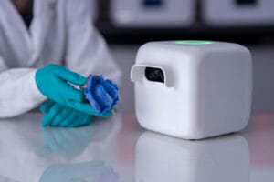
The Cubit360™ scanner from Mimetrik™ is revolutionising the digital dental workflow, enabling those who value speed, efficiency, and traditional techniques to access the benefits that digital dentistry has to offer. The scanner is fast and intuitive, offering six degrees of freedom for continuous, real-time scanning without mechanical delays. This means that, once an analogue impression is taken, it can be scanned immediately and shared with the dental lab electronically without the need to post the impression.
So, to summarise, accuracy is absolutely crucial when carrying out dental scans, often meaning that errors during these processes, caused by issues such as patient movement, result in the need for retakes. By selecting the most appropriate method for each use case and working with patients to ensure they are informed of the requirements for a successful outcome, clinicians can minimise the number of imaging errors and resulting artefacts.

For more information about Mimetrik, please visit https://mimetrik.tech/
[i] Spin-Neto, R., Costa, C., Salgado, D.M., Zambrana, N.R., Gotfredsen, E. and Wenzel, A., 2018. Patient movement characteristics and the impact on CBCT image quality and interpretability. Dentomaxillofacial Radiology, 47(1), p.20170216. https://pmc.ncbi.nlm.nih.gov/articles/PMC5965740/
[ii] Almotairi, Y. A., Almutairi, T. O., Almutairi, F. H., Al Otaibi, E. S., Almutairi, W. A., Ishaq, G. F. and Almotairi, Y. A. (2024) “Strategies for minimizing repeated exposures in diagnostic radiography”, International Journal Of Community Medicine And Public Health, 11(9), pp. 3697–3701. doi: 10.18203/2394-6040.ijcmph20242300.
[iii] Limones, A., Çakmak, G., Fonseca, M., Roccuzzo, A., Cobo-Vázquez, C., Gómez-Polo, M. and Molinero-Mourelle, P., 2025. Impact of scanning interruptions on accuracy of implant-supported full-arch scans: An in-vitro pilot study. Journal of dentistry, 153, p.105503. https://www.sciencedirect.com/science/article/pii/S0300571224006730
[iv] Fratila AM, Saceleanu A, Arcas VC, Fratila N, Earar K. Enhancing Intraoral Scanning Accuracy: From the Influencing Factors to a Procedural Guideline. Journal of Clinical Medicine. 2025; 14(10):3562. https://doi.org/10.3390/jcm14103562
[v] Clozza, E., 2024. Intraoral scanning and dental photogrammetry for full‐arch implant‐supported prosthesis: A technique. Clinical Advances in Periodontics, 14(4), pp.244-249.


